THE OVARIES
The ovaries possess both gametogenic and endocrine functions. Each ovary is a solid, basically ellipsoidal body, although commonly made irregular by the projection from the surface of large follicles and corpora lutea (Figure 5-55, A-F).
The irregularity is naturally greatest in polytocous species, in which follicles ripen in batches. The ovaries are much smaller than the testes of conspecific males but, like these, bear no constant proportion to body size. Those of the mare are relatively large and also peculiar in being kidney-shaped. Ovaries are usually found in the dorsal part of the abdomen, close to the tips of the horns of the uterus, as they do not shift far from their place of development. This migration, generally modest, occurs in the absence of any apparent endocrine influence: it is most considerable in ruminants in which the ovaries come to lie close by the pelvic inlet. Each ovary is suspended within the cranial part (mesovarium) of the broad ligament, the common suspension of the female reproductive tract.A section through the ovary of a mature animal shows it to consist of a central looser and more vascular part contained within a denser shell. The parenchymatous zone (cortex) is bounded by a tunica albuginea directly below the peritoneum and is strewn with follicles in various stages of development and regression. Each follicle contains a single ovum; the stages through which it passes are shown in schematic fashion in Figure 5-56. The rapid enlargement undergone by those follicles selected to come to maturity in the current cycle is mainly due to the accumulation of the fluid by which the ova are swept out on ovulation. The cavity within the ruptured follicle, though it may initially fill with blood, is soon occupied by hypertrophy of the granulosa and theca cells that originally lined the space. This produces a solid body, known as the corpus luteum (yellow body) on account of its color (Figure 5-55, E).
Corpora lutea are transient structures that wax and wane between one estrous period and the next (assuming pregnancy does not ensue) (Figure 5-57, A-C). Degeneration of the corpora lutea is characterized by vacuolization of the cytoplasm of the luteal cells due to lipid accumulation and nuclear shrinkage. Although transient, they are important as the source of progesterone, just as the ripening follicles are the source of estrogen. Corpora lutea finally regress and are replaced by connective tissue scars, corpora albicantes (white bodies). The alternation in the levels of estrogen and progesterone determines the changes in the behavior pattern and in the morphology and activity of the reproductive tract.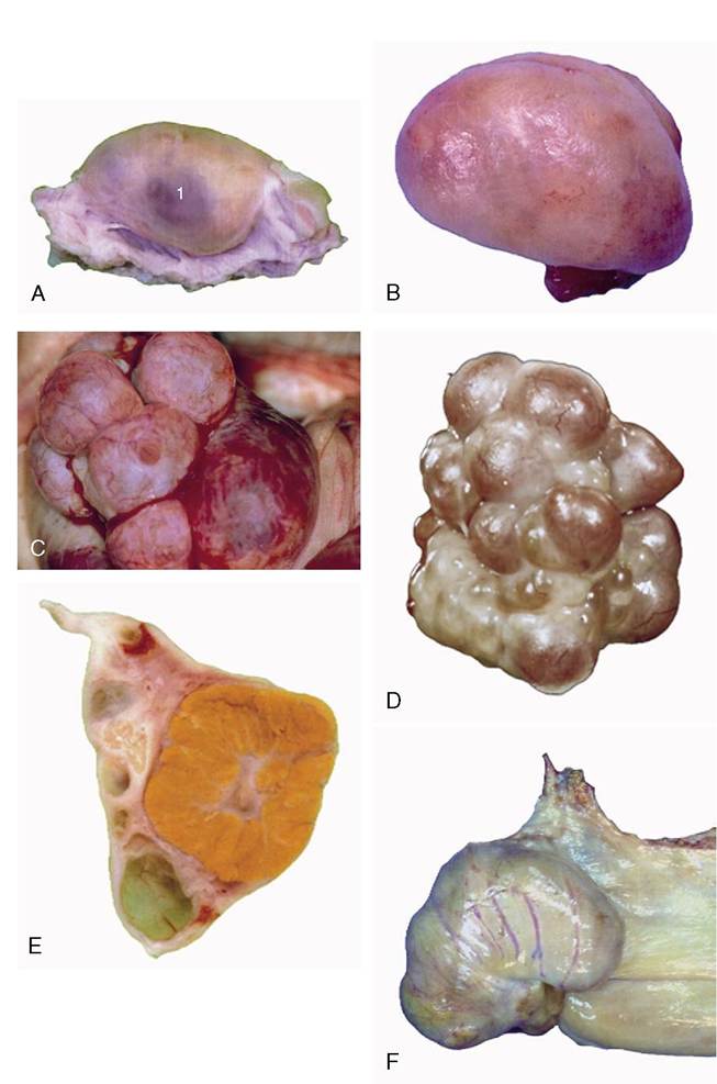
Figure 5-55 Specific and functional variations in ovarian morphology. A, Ovary of a cow (monotocous). B, Ovary of a bitch in a quiet stage. C, Ovary of a bitch exhibiting several mature follicles. D, Ovary of sow (polytocous) exhibiting mature follicles. E, Sectioned ovary of a cow containing a large corpus luteum. F, Ovary of a mare, with ovulation fossa. 1, Mature follicle.