THE UTERINE TUBES
The uterine tubes are narrow and generally very flexuous. They capture the ova released from the ovaries and convey them toward the uterus; because they also convey the sperm in their ascent, fertilization normally occurs within the tubes.
The free cranial extremity takes the form of a thinwalled funnel (infundibulum; Figure 5-58/2) placed close to the cranial pole of the ovary. The free edge of the funnel is ragged, and the tags (fimbriae) come into contact with and sometimes adhere to the surface of the ovary. A small (abdominal) orifice in the depth of the funnel leads to the longer tubular part that is divided into two more or less equal segments. The proximal one, known as the ampulla, is followed by the more convo-
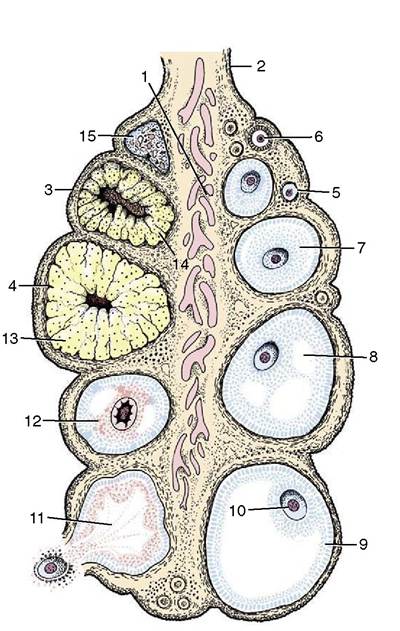
Figure 5-56 Schematic representation of the different functional stages in ovarian activity. 1, Medulla; 2, mesovarium; 3, surface epithelium; 4, tunica albuginea (poorly developed); 5, primordial follicle; 6, primary follicle; 7, secondary follicle; 8, early tertiary follicle; 9, mature follicle; 10, oocyte; 11, ruptured follicle; 12, atretic follicle; 13, corpus luteum; 14, atretic corpus luteum; 15, corpus albicans.
luted and narrower isthmus, but the distinction between these segments is not equally obvious in all species or at all phases of the cycle (Figure 5-58/3,4). The isthmus joins the apex of the horn of the uterus at the uterotubal (salpingouterine) junction, a region of very variable appearance. The junction is gradual in ruminants and pigs and abrupt in horses and carnivores; indeed, in the mare and to a lesser extent also in the bitch and cat, the terminal part of the tube is thrust into the apex of the horn to raise a small papilla perforated by the (uterine) orifice of the tube. Too much should not be made of these differences as, regardless of its appearance, the junction always represents a real barrier, impeding both the ascent of sperm and the descent of ova. The tube wall consists of external serosal, middle muscular, and internal mucosal tunics. The mucosa is folded longitudinally along its whole length from infundibulum to isthmus; secondary and even tertiary folds reduce the lumen of the ampulla to a series of narrow branching clefts. The tube is carried in a side-fold (mesosalpinx) of the part of the broad ligament that supports the ovary.