THE OVARY AND UTERINE TUBE
The ovary is a firm, rather irregular ovoid body, small (4 ? 2.5 ? 1.5 cm) in relation to body size. Joined to the body wall and to the reproductive tract by inclusion in the broad ligament, it is related to the ventral part of the shaft of the ilium, level with the bifurcation of the uterus.
Follicles and corpora lutea may project from any part of the surface (Figure 29-13).The largest follicles attain a diameter of 2 cm, but even those as small as 5 mm in diameter may be detected on palpation per rectum. Because the estrus cycle is short (generally 21 days), follicles and corpora lutea of some size may be present together.
The uterine tube is rather long, but its flexuous course brings its beginning and end close together (Figure 29-14, A-B). The thin-walled infundibulum lies over the lateral wall of the ovary in the free margin of the mesosalpinx. The succeeding, narrower part of the tube winds within the lateral wall of the ovarian bursa
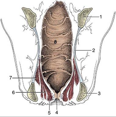
Figure 29-6 Dorsal section of the bovine rectum and adjacent structures. Note especially the topography of the pelvic diaphragm (6, 7). 1, Shaft of ilium; 2, sacrosciatic ligament; 3, ischial tuber; 4, anus; 5, external anal sphincter; 6, levator ani; 7, coccygeus; 8, rectum.
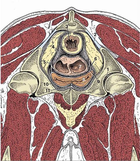
Figure 29-7 Transverse section of the pelvis of a cow at the level of the hip joint (cranial surface). Note the large amount of retroperitoneal fat in the pelvis. (See Figure 29-11 for the level of this section.) 1, Hip joint; 2, sacrosciatic ligament; 3, rectum; 4, rectogenital pouch; 5, broad ligament of uterus; 6, lateral ligament of bladder; 7, uterus sectioned where the two horns are conjoined; 8, bladder; 9, vesiocogeni- tal pouch; 10, pubovesical pouch; 11, median ligament of bladder.
to reach the tip of the uterine horn. It is divided into ampulla and isthmus, approximately in the ratio of 2:1, but the distinction is only obvious at certain stages of the cycle. The transition of isthmus to horn is gradual and marked by muscular thickening.
Apart from features associated with the frequency of twin and multiple pregnancies, the ovaries as well as the tubes of sheep and goats are very similar to those of cows.