THE UTERUS
At first sight, the uterus appears to consist of a relatively long body succeeded by two divergent, tapering horns coiled ventrally on themselves (Figure 29-15).
This impression is misleading.
Most of the apparent body is furnished by the two horns lying side by side within shared serosal and muscular coats, which is an arrangement suggested by a dorsal groove that becomes more pronounced toward the bifurcation. Where the horns diverge, the superficial tissues initially bridge the gap, forming short dorsal and ventral intercornual ligaments (Figure 29-14Z√) that bound a small pocket conveniently arranged to allow the organ to be fixed by a finger during rectal examinations. The tight winding of the horns is not constant but results from stimulation of the muscle of the organ and of the broad ligament. The stimulus is provided by handling, which explains why the form of the uterus appears to become more definite and its consistency firmer in the course of a rectal examination. The effect is most noticeable during estrus.The firmness of the cervix permits recognition of the caudal limit of the body when the intact organ is handled, but there is nothing to indicate its cranial limit. It is surprising to discover, when the organ is laid open, that the body is a mere 3 cm in length. Each horn measures 35 cm or so, of which about one third is incorporated in the “pseudobody”; the cervix measures 8 to 10 cm.
The most characteristic feature of the interior is the presence of the caruncles, the attachment sites of the fetal membranes in pregnancy. About forty of these are arranged in four more or less regular rows in the wider parts of the horn, reducing to a double line toward the tip.
The cervix begins at the constriction of the internal uterine ostium, beyond which the passage is occluded by the interlocking of projections from the walls; these consist of three or four circular folds in virgin animals,
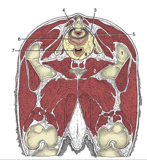
Figure 29-8 Transverse section of the bovine pelvis at the level of the first caudal vertebra (cranial surface). The section passes through the obturator foramina.
Note that the peritoneum covers only the dorsal surface of the vagina; the lateral and ventral surfaces are retroperitoneal at this level. (See Figure 29-11 for the level of this section.) 1, Greater trochanter; 2, obturator foramen; 3, sacrosciatic ligament; 4, rectum; 5, rectogenital pouch; 6, vagina; 7, neck of bladder; 8, retroperitoneal fat.but these become broken and irregular in multipara. The most caudal fold projects into the vagina, where it is surrounded by an annular fornix. The cervical mucosa also shows longitudinal folds that, on reaching the external ostium, radiate in a fashion recalling the segments of an orange (Figure 29-16, A-B). If parturition is disregarded, the cervix is most easily passed by an instrument at estrus, but the difficulties experienced at other times can be overcome and indeed must be overcome to allow embryo transfer.
Most features that distinguish the uterus of the small ruminants are of little practical importance. The free surfaces of the caruncles are concave, most obviously in the ewe (Figure 29-17). Certain features of the cervix are more significant. Many irregular, originally circular folds project into the lumen, fitting closely together; the last one is sunken into a recess of the vaginal wall. In combination, these features make catheterization of the uterus very difficult if not impossible at most stages of the cycle.
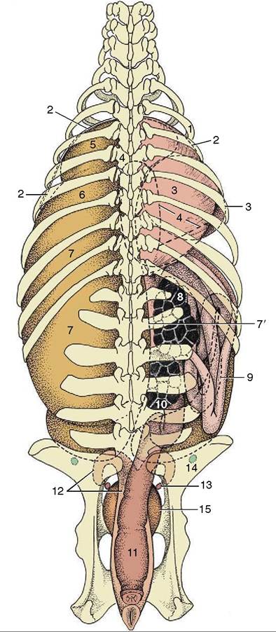
Figure 29-9 Relationship of the principal abdominal and pelvic organs to the bovine skeleton, dorsal view. 1, Sixth rib; 2, cranial extent of diaphragm; 3, omasum, most of it covered by the liver; 4, outline of abomasum; 5, reticulum; 6, atrium ruminis; 7, dorsal sac; 7’, right face of rumen; 8, right kidney;
9, descending duodenum; ventral to it is the intestinal mass;
10, left kidney; 11, rectum; 12, uterus; 13, ovary; 14, lateral iliac lymph node; 15, bladder.