The Ovary and Uterine Tube
The ovary is a firm, rather irregular ovoid body, small (4 ? 2.5 ? 1.5 cm) in relation to body size. Joined to the body wall and to the reproductive tract by inclusion in the broad ligament, it is related to the ventral part of the shaft of the ilium, level with the bifurcation of the uterus.
Follicles and corpora lutea may project from any part of the surface (Figs. 29.12 and 29.13).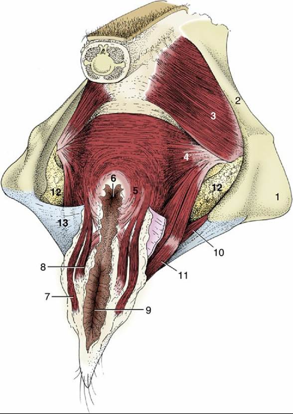
FIG. 29.10 The perineal muscles of a cow. 1, Ischial tuber; 2, sacroscιatιc ligament; 3, coccygeus; 4, levator ani; 5, external anal sphincter; 6, anus; 7, retractor clitoridis; 8, constrictor vulvae; 9, vulva; 10, urogenital diaphragm; 11, constrictor vestibuli; 12, fat in ischiorectal fossa; 13, perineal fascia (partly removed on the right side).
The largest follicles attain a diameter of 2 cm, but even those as small as 5 mm in diameter may be detected on palpation per rectum. Because the estrus cycle is short (generally 21 days), follicles and corpora lutea of some size may be present together.
Ovarian follicular cysts affect 10% of dairy cattle. Because cows are generally infertile until the cyst is treated, it increases the inter-calving interval and causes significant economic losses to the dairy industry. There is some evidence as to the heritability of the cysts.
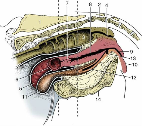
FIG. 29.11 Median section of the bovine pelvis. The two vertical broken lines indicate the levels of the transverse sections in Figs. 29.7 (left line) and 29.8 (right line). The position of the obturator foramen is indicated by a broken outline. 1, Sacrum; 2, first caudal vertebra; 3, rectum; 4, anal canal; 5, right uterine horn; 6, left uterine horn, mostly removed; 7, cervix; 8, vagina; 9, vestibule; 10, vulva; 11, bladder; 12, urethra; 13, suburethral diverticulum; 14, symphysis.
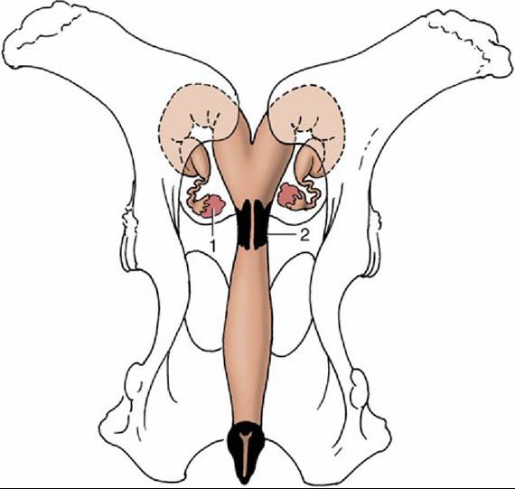
FIG.
29.12 Dorsal view of the bony pelvis and related (nongravid) bovine reproductive organs. Note the position of the ovaries in relation to the pecten pubis. 1, Ovary; 2, cervix.The uterine tube is rather long, but its flexuous course brings its beginning and end close together (Fig. 29.14A and B). The thin-walled infundibulum lies over the lateral wall of the ovary in the free margin of the mesosalpinx. The succeeding, narrower part of the tube winds within the lateral wall of the ovarian bursa to reach the tip of the uterine horn. It is divided into ampulla and isthmus, approximately in the ratio of 2:1, but the distinction is only obvious at certain stages of the cycle. The transition of isthmus to horn is gradual and marked by muscular thickening.
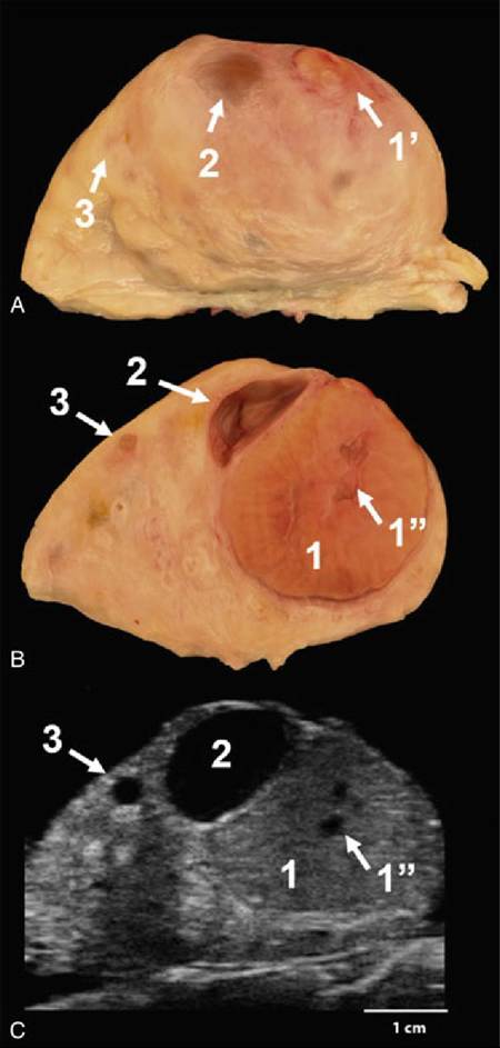
FIG. 29.13 Images showing the surface (A), a longitudinal cut view (B), and an ultrasound image (C) of the bovine ovary. Only a small part of the fully developed corpus luteum (1,) projects above the surface while the majority of the glandular tissue structure (1) is embedded within the ovary. Irregular-shaped cavities (1”) are common in the corpus luteum and easily detectable by ultrasound imaging. Note a large (2) and a small (3) antral follicle in the cortex of the ovary. Fluid-filled antrum appears as an anechoic (dark) structure in ultrasound images.
Apart from features associated with the frequency of twin and multiple pregnancies, the ovaries as well as the tubes of sheep and goats are very similar to those of cows.