» The Passive Stay Apparatus
The caudal end of the trunk rests on the head of the femur. A vertical line dropped from the center of the support passes caudal to the stifle joint and cranial to the hock, fetlock, and pastern joints before intersecting the hoof (Fig.
24.7A∣ arrow). If unsupported, the bony column of the hindlimb would collapse by flexion of the stifle and hock and overextension of the fetlock and pastern joints. The tendons and ligaments of the passive stay apparatus enable the horse to prevent this collapse using only a minimum of muscular effort.The supportive mechanisms below the hock are very similar to those of the forelimb (pp. 602606). However, the accessory ligament of the deep digital flexor tendon, which arises from the caudal aspect of the hock, is weak and occasionally absent. This is compensated by the firm, intermediate attachment of the superficial digital flexor tendon to the point of the hock, which is broadly comparable in function to the accessory ligament of the corresponding tendon of the forelimb. The part of the superficial flexor tendon between its attachments proximal and distal to the fetlock joint is tensed when weight is on the limb and assists the interosseous muscle in supporting the fetlock.
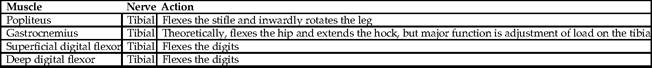
» TABLE 24-5
The Caudal Muscles of the Leg
Fixation of the stifle and hock joints depends on the locking mechanism of the former joint and the existence of the so-called reciprocal mechanism, which associates the movements of the two joints. For the horse to "lock" the stifle, the patella is first brought into the resting position (by extending the joint) and then fixed by being rotated medially through about 15 degrees (Fig. 24.7E∣αrrow). This hooks the parapatellar cartilage and medial patellar ligament securely over the protuberance of the medial trochlear ridge (Fig.
24.7∣17) and places the medial ligament more caudally, as much as 2 cm behind the crest of the medial ridge, than before. The patella now firmly resists displacement, and a larger part of the body weight can be lowered onto the locked joint, which enables the other hindlimb to be rested with only the toe of the hoof on the ground. The "unlocking" is effected quite briskly with lateral rotation of the patella to snap it back into its usual place.The reciprocal mechanism is provided by two tendinous cords—the peroneus tertius and the superficial digital flexor—that pass between the distal end of the femur and the hock, one on the cranial and the other on the caudal aspect of the tibia (Fig. 24.7∣7 and 9). (Fig. 24.16A demonstrates the result of the rupture of the peroneus tertius.) These ensure that the two joints flex or extend in unison. However, some looseness in the system renders it unnecessary for the angular changes at the two joints to be exactly the same, especially during fast gaits when large forces must be absorbed by the tendons.
When the stifle is locked, the weight of the hindquarters tends to flex the hock joint; this is opposed by tension in the superficial flexor caudal to the tibia. The peroneus tertius is not involved at this time, and it seems that it is superfluous in the animal standing quietly.
The stifle joint is fully locked only when the horse takes most of the weight on that limb and rests the other on the toe of the hoof. It should be emphasized that although the arrangement conserves energy, it does not eliminate muscular effort; every few minutes the animal shifts its support from one side to the other as muscles tire or, perhaps, as tension in the passive supporting structures becomes uncomfortable.
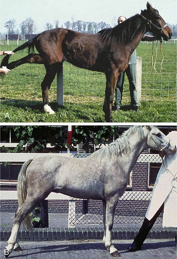
FIG. 24.16 (A) Rupture of peroneus tertius. (B) Locked patella.
Sometimes a neuromuscular disorder makes unlocking of the stifle difficult or even impossible (Fig.
24.16B). A temporary "lock" may be broken by startling a horse into sudden movement; a persistent "lock" may be alleviated by surgical section of the medial patellar ligament to break the retention loop (Fig. 24.7B/1'). The operation is easily and safely performed because a considerable thickness of fat lies deep to the ligament, protecting the synovial membrane.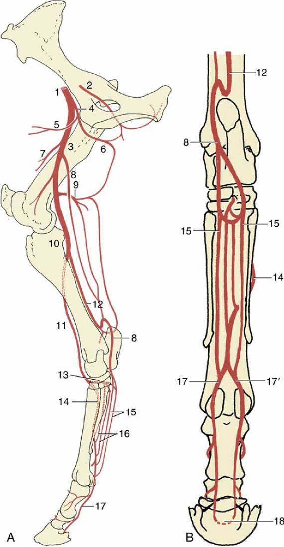
FIG. 24.17 The principal arteries (aa.) of the right hindlimb: (A) medial view; (B) caudal view. 1, External iliac artery (a.); 2, obturator a.; 3, femoral a.; 4, deep femoral a.; 5, pudendoepigastric trunk; 6, medial circumflex femoral a.; 7, lateral circumflex femoral a.; 8, saphenous a.; 9, caudal femoral a.; 10, popliteal a.; 11, cranial tibial a.; 12, caudal tibial a.; 13, perforating tarsal a.; 14, dorsal metatarsal a.; 15, medial and lateral plantar aa.; 16, medial and lateral plantar metatarsal aa.; 17 and 17', medial and lateral digital aa., respectively; 18, terminal arch, anastomosis of digital aa. within the distal phalanx.