THE PELVIC REPRODUCTIVE ORGANS
The constituents of the spermatic cord disperse at the vaginal ring, from whence the deferent duct may be traced over the dorsal surface of the bladder. It passes under the body of the prostate to reach the urethra, and in the last part of its course it is combined with the duct of the vesicular gland in a very short common passage.
The subterminal stretch (ca. 10 to 12 cm) lies beside its fellow in the genital fold; the wall of this part is swollen to form the cylindrical ampulla or ampullary gland. A median vestige of the fused paramesonephric ducts is sometimes present between the two ampullae (Figure 29-29).The urethra runs over the pelvic floor from the bladder (Figure 29-30) and leaves the pelvic cavity by bending around the ischial arch. Level with the arch, the lumen presents a dorsal diverticulum guarded at its entrance by a mucosal flap. The flap splits at its caudal extremity into two folds that constrict the urethral lumen by attaching to the walls. The tip of a catheter almost inevitably engages in this diverticulum, which makes catheterization of the bladder impossible if surgical access to the urethra is not gained first. (Even without the diverticulum, the sigmoid flexure of the penis presents a formidable complication.)
The pelvic urethra is encircled by the striated urethral muscle, completed dorsally by a stout aponeurotic plate. A thin sleeve of spongy tissue directly surrounds the lumen; when followed caudally, it expands to form the bulb of the penis. The penile urethra is narrower, especially at the sigmoid flexure, where calculi most often lodge, particularly in castrated animals.
The vesicular glands are very large (10 ? 3 to 15 ? 5 cm) and contribute the bulk of the seminal fluid. They are flexed on themselves, grossly lobulated with narrow branching lumina, and lie within the genital folds, mainly lateral to the ampullary glands (Figure 29-29, A-B).
The prostate of the bull consists of a disseminate part stretching along the length of the urethra, largely dorsal to the lumen and diminishing in thickness when followed caudally, and a compact part (body) consisting of paired lobes that have broken through the urethral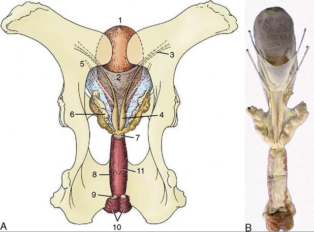
Figure 29-29 Dorsal view of the bull's pelvis and related urogenital organs. A, Schema. 1, Bladder; 2, genital fold; 3, right deferent duct; 4, ampulla of deferent duct; 5, left ureter; 6, vesicular gland; 7, body of prostate; 8, urethralis (surrounding urethra); 9, bulbourethral gland; 10, bulbospongiosus; 11, caudal extent of the rectogenital pouch (broken line). B, Specimen.
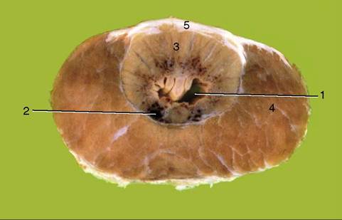
Figure 29-30 Transverse section of the bovine pelvic urethra immediately caudal to the body of the prostate. 1, Urethra; 2, spongy tissue (stratum spongiosum); 3, disseminate part of prostate; 4, urethralis; 5, dorsal aponeurosis of urethralis.
aponeurosis and together form a bar lying across the first part of the urethra (4 ? 1 cm).
The small bulbourethral glands, located by the ischial arch, are flattened and covered by the bulbospongiosus muscle (Figure 29-29, B). Their watery secretion is discharged into the diverticulum and flushes the urethra in advance of the main ejaculate.
Apart from the body of the prostate, which is specific to the bull, the pelvic reproductive glands are very similar in the three domestic ruminants.
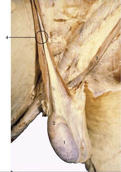
Figure 29-31 Scrotum opened, and testis and epididymis exposed. Note tortuous veins on surface of the testis. 1, Testis; 2, epididymis; 3, retractor penis muscle; 4, spermatic cord.
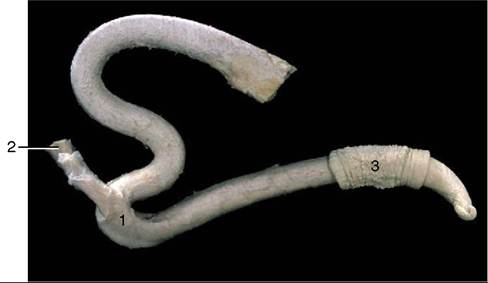
Figure 29-32 The fibroelastic bovine penis and its retractor muscle. 1, Sigmoid flexure; 2, retractor penis muscle; 3, preputial skin.