THE PENIS AND PREPUCE
The penis of an adult bull is almost 1 m long, but about a quarter of its length is taken up by the sigmoid flexure located above and behind the scrotum (Figures 29-31, 29-32, and 29-33).
Being of the fibroelastic type, it is relatively rigid at all times. The rodlike, laterally compressed crura are almost surrounded by the powerful ischiocavernosus muscles and contain more generous cavernous spaces than are present in other parts of the organ. The construction of the body of the penis is not immediately evident because its constituents, the crura and the urethra, are enclosed within a common tunica albuginea (Figure 29-34). Paired ligaments suspend the caudal part of the body from the symphysial tendon; their occasional rupture causes the penis to sag. The extremity of the quiescent penis is capped by a cushion of softer tissue, forming an asymmetrical, ventrally bent, and slightly spiraled glans that is contained within the caudal part of the prepuce. The glans exhibits a raphe or seam over its right aspect; the urethra follows this to open on the summit of a low process (Figure 29-35).
The prepuce shows the usual disposition and encloses a cavity that is both long and narrow. The prepuce droops behind the umbilicus, most obviously in beef bulls, which makes it vulnerable to injury by sharp grasses.
The penis obtains its blood supply from branches of the internal pudendal artery that are detached within the pelvis. One, the artery of the bulb, supplies the bulb and corpus spongiosum; a second, the deep artery of the penis, supplies the crus; and a third, the dorsal artery, travels along the upper border to reach the glans, detaching twigs to the prepuce en route. All three are accompanied by satellite veins that drain both the tissues and the blood spaces within the spongy and
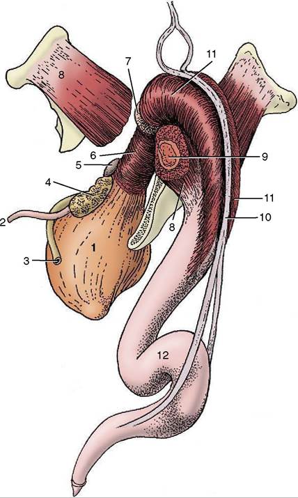
Figure 29-33 The bovine penis and its muscles; caudola- teral view.
1, Bladder; 2, ureter; 3, deferent duct; 4, vesicular gland; 5, body of prostate; 6, urethralis; 7, bulbourethral gland; 8, ischiocavernosus; 9, crus of penis (in transverse section); 10, retractor penis; 11, bulbospongiosus; 12, sigmoid flexure.cavernous bodies. The crura and corpus cavernosum constitute a single unit into which blood is transferred during erection. Venous blood leaving this unit reaches the systemic circulation via pelvic channels. The bulb, the corpus spongiosum, and the glans form a second unit that also drains via pelvic channels but possesses an additional more cranial outlet. Consequently, drainage of the spongiosus system is not completely arrested by contraction of the bulbospongiosus.
The paired dorsal nerves, which run with the dorsal arteries, overlap in their distribution. Since stimulation of the apex of the penis is necessary for the attainment of full erection, the integrity of these nerves is essential for reproductive competence. The preputial skin, including that over the penis, is supplied from the first two lumbar, the genitofemoral, and the pudendal nerves.

Figure 29-34 Cast of the cavernous spaces of the bovine penis (A) and transverse sections caudal (B) and cranial (C) to the sigmoid flexure. 1, Corpus cavernosum; 2, corpus spongiosum; 3, urethra; 4, tunica albuginea.
Cranial preputial muscles that arise in the xiphoid region and insert beside and behind the preputial orifice are able to draw the prepuce craniodorsally, which constricts its orifice. Anomalies of these muscles may prevent protrusion or impair the return of the penis to the prepuce. Caudal preputial muscles of inconstant occurrence appear to have little significance.
The usual suite of muscles is associated with the penis (see Figure 29-33). The well-developed retractor penis possesses particular interest as it must relax to allow exposure of the penis for examination or treatment.
It arises from the caudal vertebrae, passes to the side of the rectum, and reaches the penis at the second bend of the flexure; some fibers attach here, but others continue to more distal and diffuse insertions. The local contractions of the retractor that help maintain the flexure are controlled by a sympathetic innervation that is conveyed within the pudendal and caudal rectal nerves; these must be blocked to allow withdrawal of the penis for examination. The administration of an antiadrenergic tranquilizer has the same effect. A low lumbar epidural block is additionally required when anesthesia is indicated.The lymphatics from the prepuce pass to the superficial inguinal node.
The penis of the small ruminants is chiefly distinguished by the length of the slender, erectile urethral process, which projects 2 to 3 cm beyond the glans in bucks and 3 to 4 cm in rams (see Figure 29-35). In former times, as in primitive societies today, amputation of the process was performed with the intention of depriving rams of their fertilizing capacity. The sheath is also relatively short in these species.