The Pelvic Reproductive Organs
The Male Urethra
The male urethra extends from an internal orifice at the bladder neck to an external orifice at the free extremity of the penis. It is thus divisible into an internal or pelvic part and an external or spongy part; here, spongy refers to the very vascular tissue that surrounds the urethra on its leaving the pelvic cavity.
The spongy part is largely incorporated within and is considered a part of the penis. The pelvic part is joined by the deferent and vesicular (or combined ejaculatory) duct(s) a short distance from its origin from the bladder; by far the greater part of the urethra thus serves to discharge both urine and semen.Although the pelvic urethra shows regional and specific variations, it consists essentially of a mucosal tube successively invested by a vascular submucosa and a muscular tunic. The mucous membrane is thrown into longitudinal folds in the inactive state. The initial part also carries a dorsal crest that continues from the urethral orifice to end in a thickening (colliculus seminalis). The colliculus displays on its sides the slitlike orifices of the deferent ducts and the much smaller openings through which the many prostatic ducts discharge (Fig. 5.50/7). Similar but more distal openings mark the entry of the ducts of other accessory glands (Fig. 5.50/8). The submucosa contains a rather inconspicuous system of connecting blood spaces that is continuous with the vastly more generous spongy investment of the second part of the urethra. The major component of the muscle coat is the striated urethralis that encircles the tube.
The urethra is embedded in fat and other connective tissues where it lies on the pelvic floor. The dorsal surface is related to the rectum and, with species differences, to various accessory reproductive glands; usually only a narrow median strip that faces directly into the rectogenital pouch is covered by peritoneum.
The urethra is easily palpated per rectum, a procedure that may stimulate rhythmic activity of its muscle.The Accessory Reproductive Glands
The full set comprises ampullary, vesicular, prostate, and bulbourethral glands, although not all of these are present in every species (Fig. 5.51). The ampullary glands have been sufficiently described.
Paired vesicular glands (Fig. 5.51/5) are present in all domestic species except the dog and cat. Each buds from the distal part of the deferent duct in the embryo, and this relationship commonly persists. In the pig the later absorption of the ejaculatory duct into the urethra causes the vesicular gland to open separately. These glands vary greatly in appearance; in the horse they are large, externally smooth, and bladder-like, resembling the human organs that were formerly known as seminal vesicles. This term is inappropriate because in most species the glands are knobby and thickwalled with rather narrow, branched lumina. The vesicular glands lie wholly or partly within the genital fold, each lateral to the corresponding deferent duct.
A prostate (Fig. 5.51/6) is present in all domestic species. In some it consists of two parts: one is diffusely spread within the wall of the pelvic urethra, and the other is a compact body placed external to the urethralis. Both parts drain by many small ducts. The small ruminants have only the diffuse or disseminate part and the horse only the compact part. The disseminate part is vestigial in the dog and cat, but the compact part is very large and globular and so well developed that it surrounds the urethra entirely (dog) or almost so (cat).
Paired bulbourethral glands (Figs. 5.51/7 and 5.52), compound tubular glands with a secretory epithelium, lie on the dorsal aspect of the urethra close to the pelvic exit. They are found in all species other than the dog (although they are vestigial in the cat). They are of moderate size in horses and ruminants but are very substantial in the pig, in which they appear as rather irregular elongated cylinders placed to each side of the urethra.
They may drain by one or by several ducts.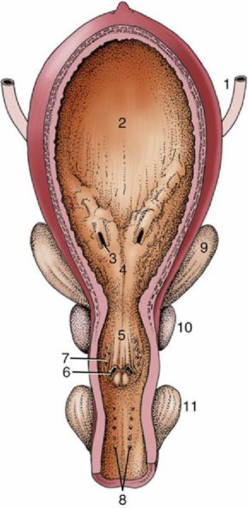
FIG. 5.50 Ventral view of the opened bladder and urethra of a stallion. 1, Ureter; 2, bladder; 3, ureteric orifice; 4, trigone of bladder; 5, urethral crest and seminal colliculus; 6, opening of ejaculatory duct; 7, multiple openings of prostatic ducts; 8, multiple openings of bulbourethral ducts; 9, vesicular gland; 10, prostate; 11, bulbourethral gland.
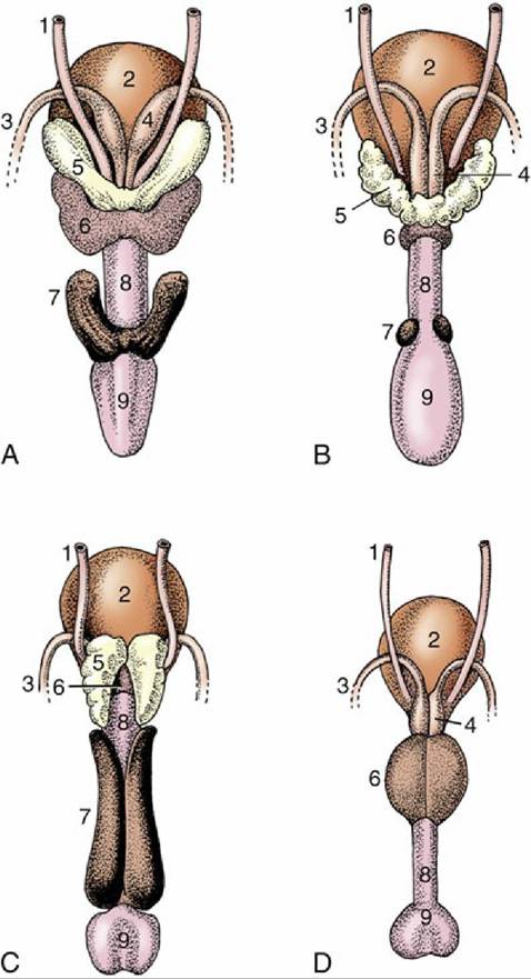
FIG. 5.51 Accessory reproductive glands of the (A) stallion, (B) bull, (C) boar, and (D) dog, dorsal view. 1, Ureter; 2, bladder; 3, deferent duct; 4, ampullary gland; 5, vesicular gland; 6, body of prostate; 7, bulbourethral gland; 8, urethra, 9, bulb of penis.
FIG. 5.52
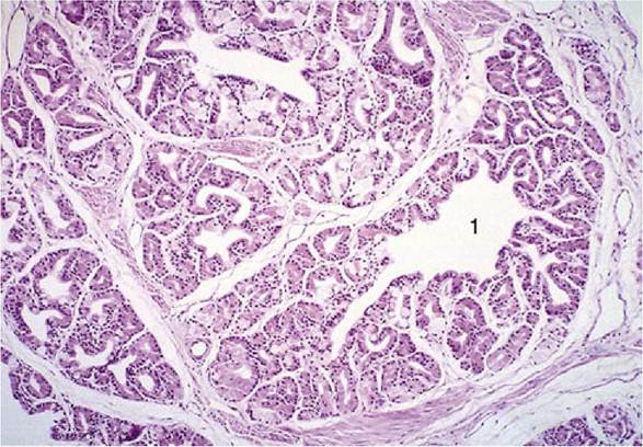
Bulbourethral gland (goat) (hematoxylin and eosin; 70?), a compound tubular gland lined with
a columnar secretory epithelium. 1, Collecting duct.
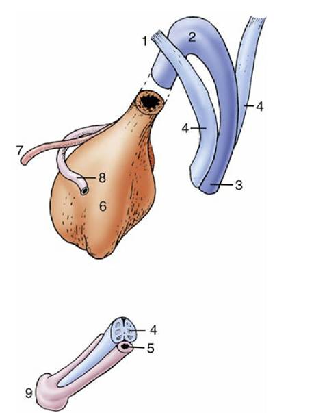
FIG. 5.53 Schematic drawing of the components that constitute the equine penis at its root (top) and at its apex (bottom). 1, Crus penis; 2, bulb; 3, corpus spongiosum; 4, corpus cavernosum; 5, urethra; 6, bladder; 7, ureter; 8, deferent duct; 9, glans.
All the larger glands possess well-developed capsules and internal septa in which much smooth muscle is present that expels the secretion at the appropriate time.