The Pelvic Reproductive Organs
The constituents of the spermatic cord disperse at the vaginal ring, from whence the deferent duct may be traced over the dorsal surface of the bladder. It passes under the body of the prostate to reach the urethra, and in the last part of its course it is combined with the duct of the vesicular gland in a very short common passage.
The subterminal stretch (≈10-12 cm) lies beside its fellow in the genital fold; the wall of this part is swollen to form the cylindrical ampulla or ampullary gland. A median vestige of the fused paramesonephric ducts is sometimes present between the two ampullae (Fig. 29.29).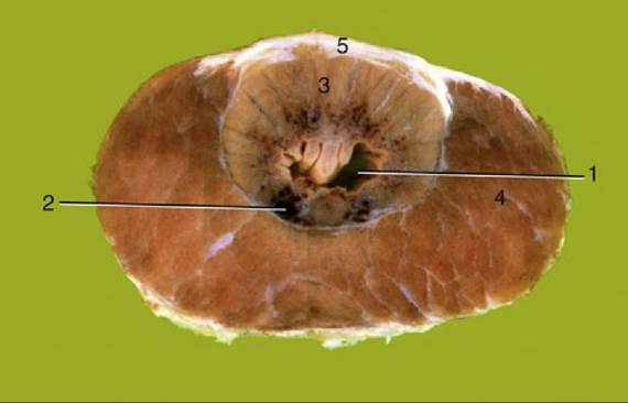
FIG. 29.30 Transverse section of the bovine pelvic urethra immediately caudal to the body of the prostate. 1, Urethra; 2, spongy tissue (stratum spongiosum); 3, disseminate part of prostate; 4, urethralis;
5, dorsal aponeurosis of urethralis.
The urethra runs over the pelvic floor from the bladder (Fig. 29.30) and leaves the pelvic cavity by bending around the ischial arch. Level with the arch, the lumen presents a dorsal diverticulum guarded at its entrance by a mucosal flap. The flap splits at its caudal extremity into two folds that constrict the urethral lumen by attaching to the walls. The tip of a catheter almost inevitably engages in this diverticulum, which makes catheterization of the bladder impossible if surgical access to the urethra is not gained first. (Even without the diverticulum, the sigmoid flexure of the penis presents a formidable complication.)
The pelvic urethra is encircled by the striated urethral muscle, completed dorsally by a stout aponeurotic plate. A thin sleeve of spongy tissue directly surrounds the lumen; when followed caudally, it expands to form the bulb of the penis. The penile urethra is narrower, especially at the sigmoid flexure, where calculi most often lodge, particularly in castrated animals.
The vesicular glands are very large (10 ? 3 to 15 ? 5 cm) and contribute the bulk of the seminal fluid. They are flexed on themselves, grossly lobulated with narrow branching lumina, and lie within the genital folds, mainly lateral to the ampullary glands (Fig. 29.29A and B). The prostate of the bull consists of a disseminate part stretching along the length of the urethra, largely dorsal to the lumen and diminishing in thickness when followed caudally, and a compact part (body) consisting of paired lobes that have broken through the urethral aponeurosis and together form a bar lying across the first part of the urethra (4 ? 1 cm).
The small bulbourethral glands, located by the ischial arch, are flattened and covered by the bulbospongiosus muscle (Fig. 29.29B). Their watery secretion is discharged into the diverticulum and flushes the urethra in advance of the main ejaculate.
Apart from the body of the prostate, which is specific to the bull, the pelvic reproductive glands are very similar in the three domestic ruminants.
The Penis and Prepuce
The penis of an adult bull is almost 1 m long, but about a quarter of its length is taken up by the sigmoid flexure located above and behind the scrotum (Figs. 29.31, 29.32, and 29.33).
The fibroelastic nature of the penis keeps it relatively rigid at all times. The rodlike, laterally compressed crura are almost surrounded by the powerful ischiocavernosus muscles and contain more generous cavernous spaces than are present in other parts of the organ. The construction of the body of the penis is not immediately evident because its constituents, the crura and the urethra, are enclosed within a common tunica albuginea (Fig. 29.34). Paired ligaments suspend the caudal part of the body from the symphyseal tendon; their occasional rupture causes the penis to sag. The extremity of the quiescent penis is capped by a cushion of softer tissue, forming an asymmetrical, ventrally bent, and slightly spiraled glans that is contained within the caudal part of the prepuce.
The glans exhibits a raphe or seam over its right aspect; the urethra follows this to open on the summit of a low process (Fig. 29.35).The prepuce shows the usual disposition and encloses a cavity that is both long and narrow. The prepuce droops behind the umbilicus, most obviously in beef bulls, which makes it vulnerable to injury by sharp grasses.
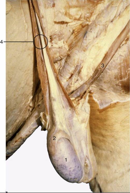
FIG. 29.31 Scrotum opened, and testis and epididymis exposed. Note tortuous veins on surface of the testis. 1, Testis; 2, epididymis; 3, retractor penis muscle; 4, spermatic cord.
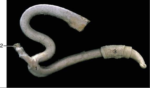
FIG. 29.32 The fibroelastic bovine penis and its retractor muscle. 1, Sigmoid flexure; 2, retractor penis muscle; 3, preputial skin.
The penis obtains its blood supply from branches of the internal pudendal artery that are detached within the pelvis. One, the artery of the bulb, supplies the bulb and corpus spongiosum; a second, the deep artery of the penis, supplies the crus; and a third, the dorsal artery, travels along the upper border to reach the glans, detaching twigs to the prepuce en route. All three are accompanied by satellite veins that drain both the tissues and the blood spaces within the spongy and cavernous bodies. The crura and corpus cavernosum constitute a single unit into which blood is transferred during erection. Venous blood leaving this unit reaches the systemic circulation via pelvic channels. The bulb, the corpus spongiosum, and the glans form a second unit that also drains via pelvic channels but possesses an additional more cranial outlet. Consequently, drainage of the spongiosus system is not completely arrested by contraction of the bulbospongiosus.
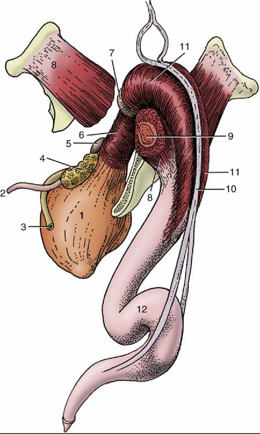
FIG. 29.33 The bovine penis and its muscles: Caudolateral view.
1, Bladder; 2, ureter; 3, deferent duct;4, vesicular gland; 5, body of prostate; 6, urethralis; 7, bulbourethral gland; 8, ischiocavernosus; 9, crus of penis (in transverse section); 10, retractor penis; 11, bulbospongiosus; 12, sigmoid flexure.
The paired dorsal nerves, which run with the dorsal arteries, overlap in their distribution. Because stimulation of the apex of the penis is necessary for the attainment of full erection, the integrity of these nerves is essential for reproductive competence. The preputial skin, including that over the penis, is supplied from the first two lumbar, the genitofemoral, and the pudendal nerves.
Cranial preputial muscles that arise in the xiphoid region and insert beside and behind the preputial orifice are able to draw the prepuce craniodorsally, which constricts its orifice. Anomalies of these muscles may prevent protrusion or impair the return of the penis to the prepuce. Caudal preputial muscles of inconstant occurrence appear to have little significance.
The usual suite of muscles is associated with the penis (Fig. 29.33). The well-developed retractor penis possesses particular interest because it must relax to allow exposure of the penis for examination or treatment. It arises from the caudal vertebrae, passes to the side of the rectum, and reaches the penis at the second bend of the flexure; some fibers attach here, but others continue to more distal and diffuse insertions. The local contractions of the retractor that help maintain the flexure are controlled by a sympathetic innervation that is conveyed within the pudendal and caudal rectal nerves; these must be blocked to allow withdrawal of the penis for examination. The administration of an antiadrenergic tranquilizer has the same effect. A low lumbar epidural block is additionally required when anesthesia is indicated.
The lymphatics from the prepuce pass to the superficial inguinal node.
The penis of the small ruminants is chiefly distinguished by the length of the slender, erectile urethral process, which projects 2 to 3 cm beyond the glans in bucks and 3 to 4 cm in rams (Fig. 29.35C and D). In former times, as in primitive societies today, amputation of the process was performed with the intention of depriving rams of their fertilizing capacity. The sheath is also relatively short in these species.