The Scrotum and Testes
The pendulous scrotum is contained between the cranial parts of the thighs and may reach the level of the hocks. A constricted neck joins it to the trunk, just caudal to the superficial inguinal ring, while its lower part is molded on the testes (Fig.
29.27). A mass of fat ("cod fat") is commonly found about the cord stump of the castrate; when present in excess, it may dilate the inguinal canal and produce a pseudohernia inguinalis. Although the rudimentary teats often found on the cranial face of the scrotum possess little intrinsic interest, their number and spacing receive attention in dairy bulls because the corresponding characters are likely to be transmitted to their female offspring. The scrotal nerve supply is diffuse; it comes from the first two lumbar, the genitofemoral, and the pudendal nerves.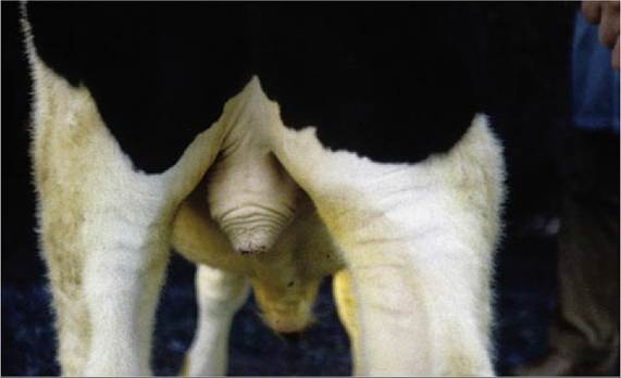
FIG. 29.27 Scrotum of bull. Musculature in tunica dartos has been contracted.
Wool covering the scrotum of the ram may cause infertility by impairing the dissipation of heat.
Each testis is ellipsoidal, is large in relation to body size (especially in the smaller ruminants), and hangs vertically in the scrotum, where it may be palpated (Fig. 29.28). It carries a large epididymis along the medial or caudomedial border that is turned to face its fellow. The epididymis is firmly attached to this border of the testis; the head extends a considerable distance down the free border, while the large, conical, and very distinctly palpable tail projects ventrally. The capsule of the testis displays a distinctive winding pattern of vessels and contains the parenchyma under slight pressure. The capsule also sends delicate partitions into the testicular tissue to form a prominent mediastinum (see Figs. 5.37 and 5.38).
After emerging from the tail, the deferent duct ascends along the medial border of the epididymis but is separated from this by the mesorchium, which is a relationship that advises a cranial approach in vasectomy operations. The duct is easily recognized on palpation as a firm, narrow strand.
The conical, dorsally tapering spermatic cord is largely composed of the exceptionally convoluted testicular artery embedded in the pampiniform plexus (see Fig. 5.43). The significance of the arteriovenous anastomoses found here remains obscure (see Fig. 5.46).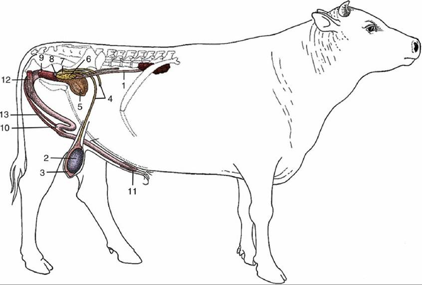
FIG. 29.28 Disposition of the urogenital organs of a bull. 1, Ureter; 2, right testis; 3, epididymis; 4, deferent duct; 5, bladder; 6, vesicular gland; 7, ampulla of deferent duct; 8, body of prostate; 9, bulbourethral gland; 10, sigmoid flexure of penis; 11, glans penis; 12, ischiocavernosus; 13, retractor penis.
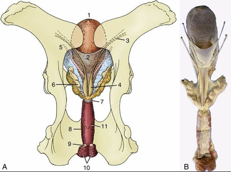
FIG. 29.29 Dorsal view of the bull's pelvis and related urogenital organs. (A) Schema. 1, Bladder; 2, genital fold; 3, right deferent duct; 4, ampulla of deferent duct; 5, left ureter; 6, vesicular gland; 7, body of prostate; 8, urethralis (surrounding urethra); 9, bulbourethral gland; 10, bulbospongiosus; 11, caudal extent of the rectogenital pouch (broken line). (B) Specimen.
Castration is an important surgery in young bulls (1-3 months of age). The castration may be performed by crushing the spermatic cord as in the Burdizzo method (closed method of castration). In the open or surgical method of castration, both of the testes are removed usually without the application of any anesthetic; however, lidocaine may be applied locally for analgesia.
The lymphatic drainage of the testis is to the medial iliac nodes; that of the scrotum is to the superficial inguinal node by the scrotal neck.