THE PENIS AND PREPUCE
The penis of the horse is composed of the usual triad of structures and is of the musculocavernous variety. The two dorsal elements, the crura penis, arise from the ischial arch, bend forward between the thighs, and soon unite in a single corpus cavernosum, which is divided in its proximal part by a median septum that reflects the compound origin (Figure 22-22, A/3).
The septum fades and finally disappears when followed toward the apex. The corpus cavernosum is somewhat compressed laterally and carries ventrally a groove into which the third erectile body, the corpus spongiosum, fits.The corpus spongiosum expands over the apex of the organ to form the distinctively shaped glans (Figure 22-22, A/1). This has a resemblance to a mushroom; the widest part, the corona, is some distance proximal to the apex, where the terminal part of the urethra protrudes into a central fossa (Figure 22-22/3). The glans is constricted to form a neck behind the corona and is then prolonged in a tapering process over the dorsal aspect of the body; this feature is not visible externally (Figure 22-21/7).
A considerable portion of the quiescent penis projects into the preputial cavity. The equine prepuce (sheath) is peculiar in being thrown into an additional fold that allows for the considerable lengthening of the penis on erection (Figure 22-22, C). The entrance (preputial ring; Figure 22-22, B/5') to this inner sleeve lies just within the preputial orifice. Sometimes as a congenital defect, the ring is unduly tight and prevents protrusion of the penis (phimosis). The condition may be corrected by section of the responsible encircling band of muscle that is included within the ring. The preputial lining contains many glands and is commonly fouled by their secretion, the smegma. An inspissated mass of this dark material—the “bean” of the penis is the stable term—commonly fills a small (urethral) sinus above the urethral process (Figure 22-22).
The penis of the horse obtains blood from the obturator and external pudendal arteries in addition to the usual internal pudendal source.
Unusually, the bulbospongiosus continues along the ventral aspect of the penis well beyond the point of incorporation of the urethra (Figure 22-21/5). The muscle, which is the direct continuation of the urethralis, bridges the ventral groove of the corpus cavernosum and on contraction compresses the corpus spongiosum (and urethra), assisting in the expulsion of urine and semen. The ischiocavernosus muscles are powerful but in no way remarkable. The smooth retractor penis muscles loop round the rectum before passing onto the ventral surface of the penis (Figure 22-21/6). They continue forward, gradually weaving through the transverse
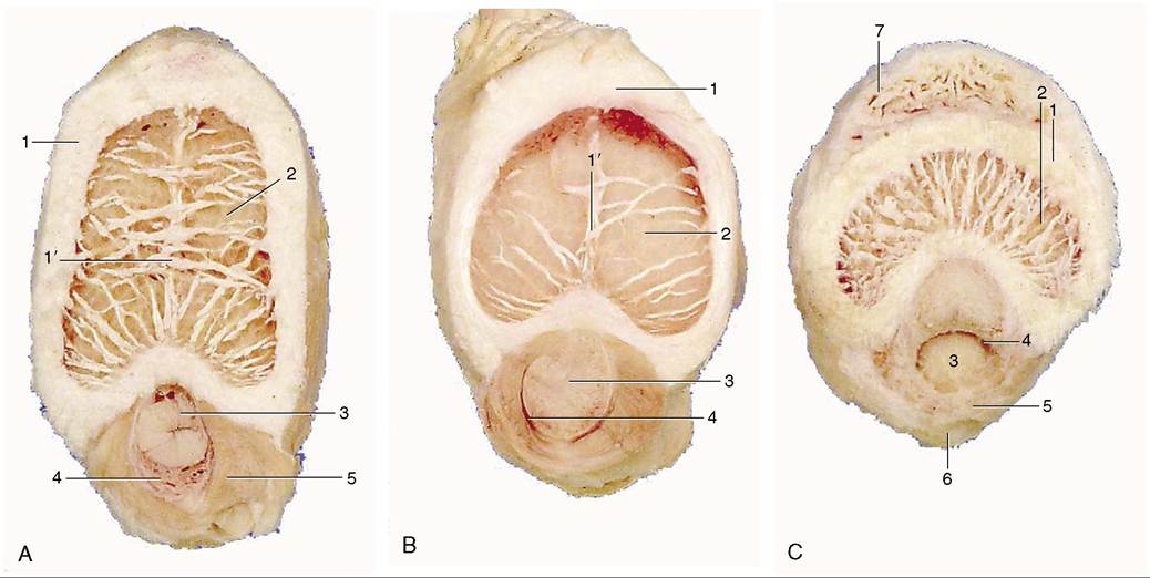
Figure 22-21 Transections of the penis, directly distal to the root (A), midshaft (B), and in its free part (C). 1, Tunica albuginea; 1', incomplete septum penis; 2, corpus cavernosum; 3, urethra; 4, corpus spongiosum; 5, bulbospongiosus; 6, retractor penis; 7, dorsal process of glans.
774" class="lazyload" data-src="/files/uch_group31/uch_pgroup304/uch_uch7232/image/image764.jpg">
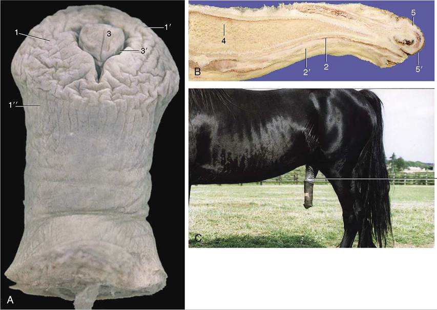
Figure 22-22 Extremity of penis exposed (A), within prepuce in median section (B), and the entire organ after dismount (C) enlarged glans penis (D). 1, Glans; 1', corona glandis; 1", collum glandis; 2, urethra; 2', corpus spongiosum; 3, urethral process within fossa glandis; 3’, urethral sinus; 4, corpus cavernosum; 5, preputial fold; 5', preputial ring; 6, prepuce, forming preputial orifice with the body wall.
fibers of the bulbospongiosus, to find attachment on the glans.
Erection
Since the penis is of the musculocavernous type, it becomes considerably engorged with blood when erect. When erection is complete, a process requiring some time and achieved by the relaxation of the helicine arteries and the pumping action of the ischiocavernosi, the organ is much enlarged in both length and girth (Figure 22-22, C).
A very considerable pressure, perhaps as much as 3700 mm Hg, is attained within the blood spaces of the corpus cavernosum, and as in other species, this occasionally results in rupture of the fibrous capsule. The ejaculate is relatively large (≈65 mL on average) and is mainly the product of the vesicular glands.Dismounting after service is often followed by a remarkable “flaring” or enlargement of the glans, in which the corona may briefly attain a diameter of 12 cm or so before it subsides. The return of the flaccid penis to the sheath is effected by the retractor muscles assisted by the smooth muscle component of the walls of the cavernosus spaces. Indeed, the resting posture of the penis is dependent on the tonus of this muscle. If this is reduced or lost—a relatively common occurrence in horses that are fatigued or in poor condition—the penis limply droops from the prepuce. It is vulnerable to injury when exposed in this way. The resistance of the muscle may also be overcome by sustained traction when it is necessary to expose the organ for clinical examination or for washing as part of routine stable hygiene.