The Pharynx
The pharynx lies wholly beneath the skull, to which the rostral third of its roof is directly applied. The remaining part of the roof and the lateral walls are enveloped by the guttural pouches (see further on).
The lumen is clearly divided into upper and lower compartments by the soft palate and the palatopharyngeal arches, which extend over the lateral walls to meet directly above the entrance to the esophagus (Fig. 18.11). The most prominent features of the nasopharynx are the flaps guarding the entrances to the auditory tubes. Each tube is about 3 cm long and is pressed against the pharyngeal wall, presenting an oblique and rather sinuous ventral free edge (Fig. 18.27A). It is stiffened by a flange of cartilage, the expansion of the medial cartilage that supports the auditory tube. The slitlike opening lateral to the flap is normally held closed but becomes patent during swallowing to equalize the pressure on the two sides of the tympanic membrane. The maneuver, which can be observed endoscopically, involves the flap swinging medially while the soft palate rises and momentarily narrows the lumen of the nasopharynx (Fig. 18.28).Endoscopy: The flap to the auditory tubes can also be elevated passively. It is a relatively simple matter to introduce an endoscope to examine, or a catheter to drain or irrigate, the guttural pouch. The entrance to the tube lies in the transverse plane of the lateral angle of the eye, which is a useful external guide to its position. The instrument encounters resistance during passage through the ventral meatus and nasopharynx. The firm support offered to its tip by the vertical lamina of the pterygoid bone is lost only a short distance rostral to the opening. Advancement of the instrument to this level generally provokes a swallowing movement when deflection of the cartilage flap facilitates entry to the pouch.
Even when performed blindly, the absence of resistance to deeper penetration indicates that the pharyngotubal opening has been successfully passed.The lower compartment of the pharynx is divided between the oropharynx and the laryngopharynx (Fig. 18.29/4 and 5). The narrow oropharynx extends between the attachment of the palatoglossal arches to the tongue and the epiglottis. Its lateral walls and floor contain much diffuse tonsillar tissue, including the long palatine tonsil (see Fig. 3.26A). The laryngopharynx is largely occupied by the projection of the larynx, and its floor is reduced to the narrow flanking piriform recesses. The laryngopharynx narrows abruptly to the origin of the esophagus.
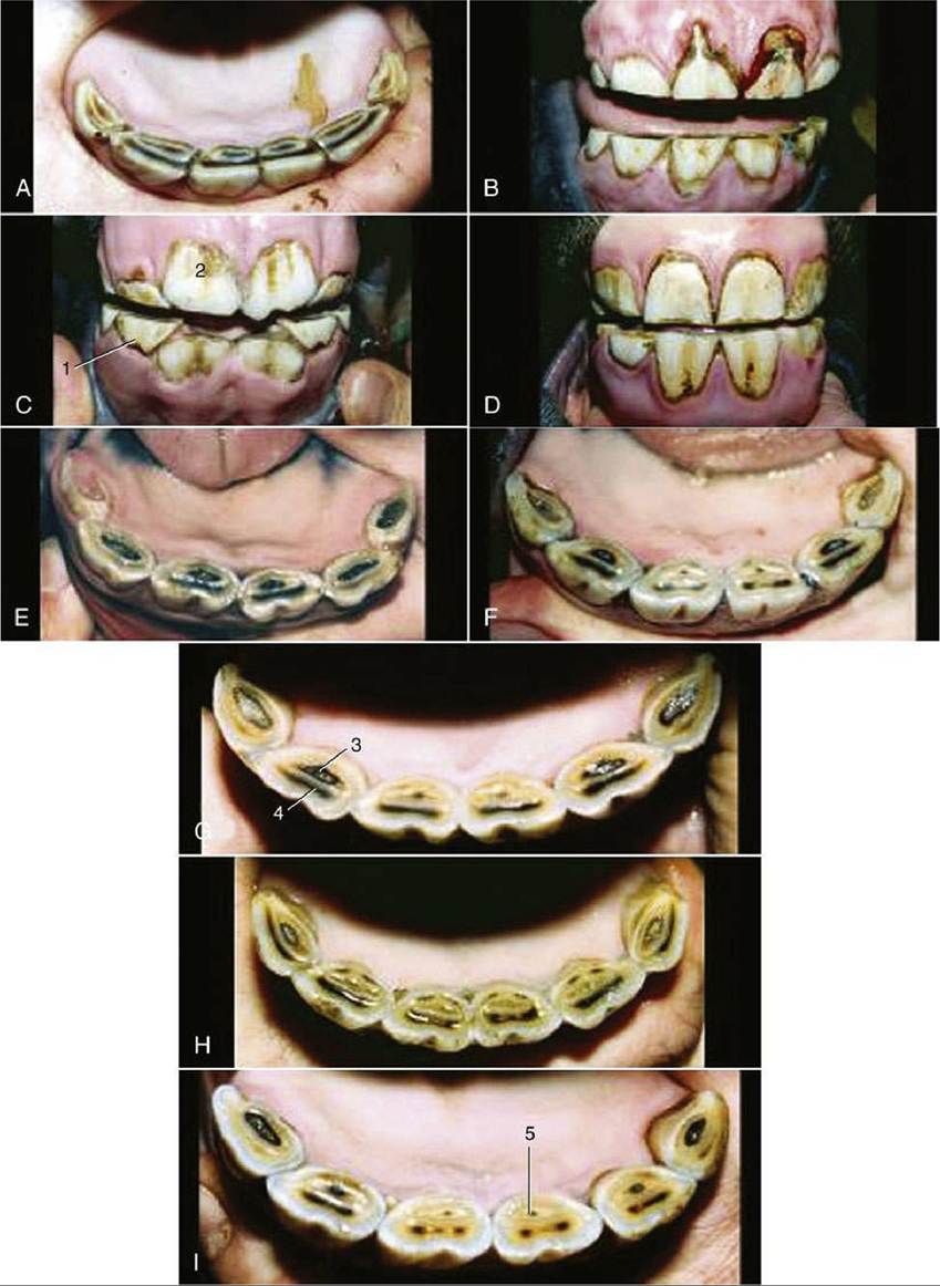
FIG. 18.24 Characteristic appearance of lower incisors of Standardbred horses of accurately known ages. (A) 112 years. (B) 212 years. (C) 3 years. (D) 4 years. (E) 5 years. (F) 6 years. (G) 7 years. (H) 8 years. (I) 9 years. 1, Deciduous teeth; 2, newly erupted I1; 3, dental cup; 4, dental star; 5, enamel spot (proximal end of infundibulum).
The structure and musculature follow the common pattern (Fig. 18.30). Difficulties in swallowing sometimes arise from malfunction of the palatine and pharyngeal muscles. The cause frequently lies in involvement of the relevant glossopharyngeal and vagus nerves in infections of the guttural pouch; because the nerves run together, they are equally susceptible (Fig. 18.26/7 and 14).
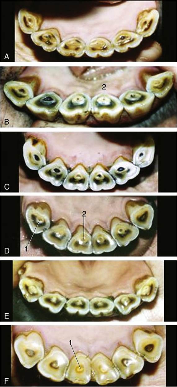
FIG. 18.25 Characteristic appearance of lower incisors of Standardbred horses of accurately known ages: (A) 11 years. (B) 12 years. (C) 14 years. (D) 16 years. (E) 17 years. (F) 20 years. Note particularity of the changes in form of the occlusal surface from round to triangular. 1, Dental star; 2, enamel spot.