The preceding chapters, which describe the physiology of movement, discuss the function of lower motor neurons, the “final common pathway" through which the central nervous system (CNS) can initiate and control movement through the contraction of skeletal muscle.
The corticospinal system and the descending brainstem motor system are described in those previous chapters as major subgroups of upper motor neurons that influence the lower motor neuron.
More medial portions of those systems coursing through the spinal cord are primarily responsible for the control of axial and proximal antigravity extensor muscles. The more lateral portions primarily control more skilled, learned, voluntary movements caused by contraction of distal flexor muscles. I his chapter describes the function of the cerebellum, part of another subgrouping of upper motor neurons critical for proper movement.The cerebellum (Latin for “little brain") is caudal to the cerebral cortex and dorsal to the brainstem (Figure 12-1). Although it constitutes only about 10% of the gross brain volume because of its highly convoluted structure, the cerebellum contains more than half of all the brain's neurons. The outer layer of cerebellar gray matter, the cerebellar cortex, has a highly regular, three-layered, histological appearance, which suggests that all cerebellar regions may perform a common underlying task. As with the cerebral cortex, the particular inputs to a given region of cerebellar cortex, and the particular output targets that it influences, in large part account for the functional differences between cerebellar regions. Two large pairs of white matter stalks, called cerebellar peduncles, primarily carry axons into the cerebellum, and a third pair of cerebellar peduncles carries axons out from the cerebellum. In addition to the cerebellar cortex and the cerebellar white matter axons entering and leaving the cortex, a group of deep cerebellar nuclei is embedded within the cerebellar white matter. The cells of these nuclei arc a principal origin of the axons leaving the cerebellum.
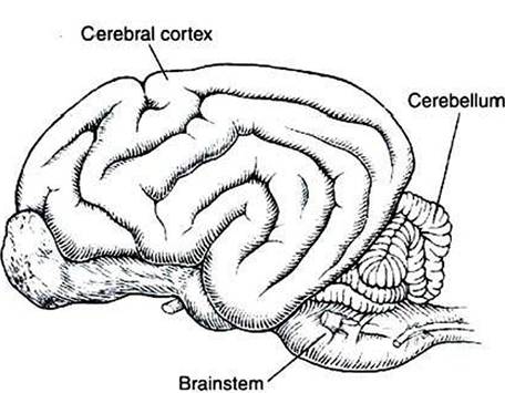
FIGURE 12-1 The cerebellum (Latin for "little brain") is caudal to the cerebral hemispheres and dorsal to the brainstem. (Redrawn from Miller ME, Christiansen GC, Evans HE: The anatomy of the dog, Philadelphia, 1964, Saunders.)
The cerebellum is not necessary for sensation or the initiation of movement. Muscle strength remains largely intact with complete destruction of the cerebellum. However, the cerebellum plays a crucial role in the timing and coordination of movement initiated by the parts of the motor system hierarchy discussed in Chapter 10. It does so by adjusting and modulating the output of the motor cortices, corticospinal tract, descending brainstem motor pathways, and spinal cord. Lesions of the cerebellum lead to major clinical deficits in the precision and grace with which movement is accomplished.