The rectum joins the anal canal ventral to the second or third caudal vertebra. Although the intraperitoneal cranial part is joined to the pelvic roof by a short mesorectum (Fig. 15.4/4),
the caudal part becomes entirely retroperitoneal once the serous covering is reflected onto the pelvic walls and the dorsal surface of the reproductive tract (bitch) or prostate (dog).
The rectum is related dorsally to the ventral muscles of the tail and certain smooth muscle bundles (rectococcygeus) that run caudally from the rectal wall to the undersurface of the tail; these bundles probably help draw the anus caudally when a column of feces descends from the colon. The ventral relations of the rectum of the bitch are the cervix and, possibly, the body of the uterus in addition to the vagina; in the male dog they are the prostate and urethra. Laterally, the rectum is bounded by the levator ani muscle and crossed by the internal pudendal vessels (Fig. 15.3) and the sciatic, pelvic, pudendal, and caudal rectal nerves. The rectum has some freedom to deviate from its usual median course because of its mesorectum and its cushioning by fat.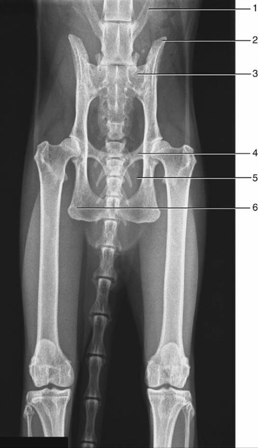
FIG. 15.2 Radiograph of the feline pelvis. 1, Transverse process of last lumbar vertebra (L7); 2, iliac crest; 3, sacrum; 4, pecten of the pubis; 5, obturator foramen; 6, ischial tuber.
The mucosa of the short (about 7 mm) initial columnar portion of the anal canal is fashioned by underlying vessels into a series of longitudinal ridges whose interdigitation helps maintain continence (Fig. 15.5). These ridges end on a line that represents the junction between the columnar intestinal epithelium and the stratified cutaneous epithelium. The outer cutaneous zone is of variable extent; the modified skin that lines this last part of the passage may be everted to appear as a purplish patch on the perineal surface, especially when defecation impends. At this time the anal orifice takes on a triangular form in place of the transverse slit generally displayed (see Fig.
10.29A).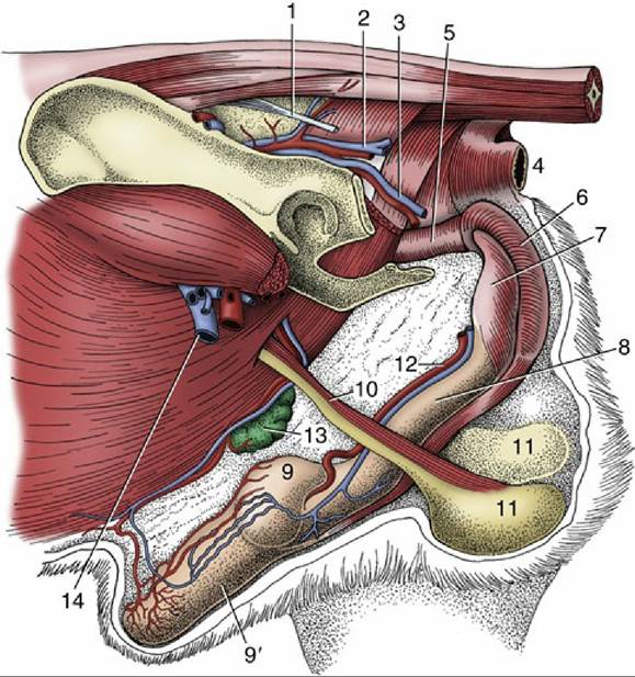
FIG. 15.3 Deep dissection of the external reproductive organs of the dog. 1, Sacrotuberous ligament; 2, caudal gluteal vessels; 3, internal pudendal vessels; 4, anus; 5, pelvic urethra; 6, bulb of penis enclosed by bulbospongiosus; 7, ischiocavernosus over left crus; 8, body of penis; 9 and 9', bulbus and pars longa glandis; 10, spermatic cord; 11, testes in scrotum; 12, dorsal artery and vein of the penis; 13, superficial inguinal lymph nodes and caudal superficial epigastric vessels; 14, femoral vessels.
Developmental errors lead to an imperforate anus, which results from the persistence of an unusually thick anal membrane, or to the absence of a longer portion of patent bowel, which results from the failure of the rectum to make proper connection with the anal pit.
All fissiped carnivores (other than bears) possess paired anal sacs (sinus paranales) enclosed between the external and internal anal sphincters. In the dog, each is about 1 cm in diameter and discharges through a short duct that opens ventrolateral to the anal orifice at the level of the anocutaneous line, concealed or exposed on the perineal surface according to the physiologic condition (see Fig. 3.47/1). In cats, the ducts of the anal sacs open on small projections some distance lateral to the anus and not at the mucocutaneous junction as in dogs. Modified sweat glands are located beneath the epithelium and discharge into the lumen of the sac. In the cats, only apocrine glands are found, but in dogs both sebaceous and apocrine sweat glands are present. Because occlusion of the duct of the anal sac is frequently encountered in dogs but is rare in cats, it is thought that the lipid component of these sebaceous secretions is responsible for the difference. The evil-smelling content of the anal sacs, normally expressed in the later stages of defecation, serves as a marker that identifies the animal to other members of its species.
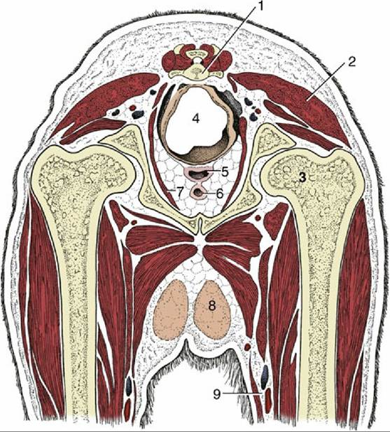
FIG. 15.4 Transverse section of the canine pelvis at the level of the hip joint. 1, Caudal vertebra; 2, superficial gluteal muscle; 3, head of femur in acetabulum; 4, rectum suspended by a short mesorectum;
5, vagina; 6, urethra; 7, levator ani; 8, inguinal mammary gland; 9, femoral artery and vein.
The impaction of anal sacs due to accumulation of secretions, generally secondary to inflammation of the sacs, is commonly seen in dogs. The anal sacs of dogs and bitches develop the malignant tumors of the apocrine glands. These tumors produce a parathormone-like hormone that raises the blood calcium levels.
The lymphatics of the anal sac drain to the sacral, hypogastric, and medial iliac lymph nodes.
There are, in addition, small anal glands within the columnar zone and much larger and more numerous circumanal or perianal glands within the cutaneous zone. In dogs, the circumanal glands are lobulated, modified sebaceous glands located in a ring about the anus, extending outward for a distance of perhaps 3 cm from the anocutaneous junction. These glands can be identified shortly after birth and increase in size throughout adult life in response to androgens. In older male dogs, slow-growing, generally benign tumors of these glands commonly develop near the anus.
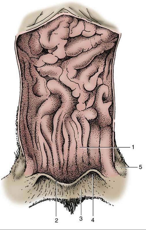
FIG. 15.5 Feline anal canal opened dorsally. 1, Columnar zone; 2, anocutaneous line; 3, cutaneous zone; 4, opening of the right anal sac; 5, right anal sac.