The Renal Pelvis and Ureter
In cattle the ureter is formed by the coming together of the short passages that lead from the calices that enclose individual renal papillae (Figs. 5.24B). In most domestic species the ureter begins in a common expansion, the renal pelvis, into which all the papillary ducts open—although in different ways in different species (Figs.
5.24). Few differences in pelvic anatomy are of practical significance. However, in the dog and cat the form of the renal pelvis obtains an importance lacking in the other species from its ready depiction in radiographs. The renal pelvis of these animals is molded on the renal crest and extends flanges dorsal and ventral to it. Each flange shows a number of local expansions or recesses that are divided from each other by projections of renal tissue (Fig. 5.29). Neighboring recesses are also separated by the interlobar vessels.The remaining tubular part of each ureter has a fairly even caliber. It mostly travels sagittally against the abdominal roof and may exhibit occasional sharp changes in direction. Each ureter bends medially upon entering the pelvic cavity to enter the genital fold in the male or the broad ligament in the female for its journey over the dorsal surface before opening into the bladder near its neck (Fig. 5.30). In the male the ureter passes dorsal to the corresponding deferent duct.
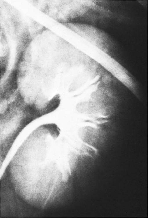
FIG. 5.29 Radiograph of the renal pelvis of a dog. Note the pelvic recesses.
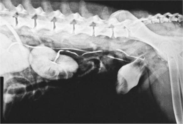
FIG. 5.30 Radiograph of the renal pelves, ureters, and bladder of a dog.
The ureter penetrates the bladder wall very obliquely, thereby guarding against reflux of urine into the ureter when the bladder fills up (Fig. 5.31). However, the angle does not prevent further filling of the bladder because the resistance is overcome by peristaltic contractions of the ureteric wall. The wall of the renal pelvis and ureter possesses an external adventitia, a well-developed middle muscularis, and an internal mucosa.