The Urinary Bladder
The distensible nature of the bladder creates variability in size, position, and relationships. When fully contracted, it is small and globular with thick walls and negligible lumen.
It is confined to the pelvic cavity in the larger species but extends into the abdomen in carnivores. The enlarged bladder is pear shaped and has a cranial vertex (apex), an intermediate body, and a caudal neck that narrows to the internal urethral orifice at the junction with the urethra. The continuing distention carries an ever-increasing portion of the bladder into the abdomen. But the neck remains fixed within the pelvis through its continuity with the urethra (Fig. 5.32/11).The initial filling of the bladder does not cause an immediate increase in internal pressure. However, considerable filling of the bladder leads to sharp rise in pressure and an urge to urinate followed by actual act of urination by most of the domestic animals. The house-trained dog may resist the urge, leading to discomfort and, later, pain as the bladder continues to fill and distend to an extent at which the apex of the bladder rests cranial to the umbilicus and creates the risk of rupture. Though the outline of the grossly distended bladder is smooth, that of the more modestly distended organ is irregular (see Fig. 5.30).
The contracted bladder in the larger species is largely retroperitoneal. However, it becomes intraperitoneal after even moderate expansion. Three folds continue this serosal covering onto the abdominal and pelvic walls (Fig. 5.33). Paired lateral vesical folds convey the round ligaments of the bladder; these vestiges of the umbilical arteries retain narrow lumina to let some blood reach the cranial part of the bladder. The third, median vesical fold, is empty in the adult, but in the fetus it supports the urachus, the constricted cranial continuation of the bladder that leaves the abdomen through the umbilical foramen before expanding externally into the allantoic sac.
The urachus and umbilical arteries rupture at birth; the urachus remains as a scar on the bladder vertex, but the umbilical arteries become the round ligaments. The folds in the adult bound the ventral pair of the several excavations into which the pelvic peritoneal cavity is divided (see Figs. 22.6 and 22.7).The constant dorsal relations of the bladder are to the uterus and vagina within the broad ligament in the female and to the deferent duct (and perhaps the vesicular glands) within the genital fold in the male. The bladder may also make indirect contact with the rectum through these folds. The ventral surface touches the pelvic and abdominal floor. Other relations of the intraabdominal part of the bladder are less predictable because of size and shape changes.
The loose attachment of the mucosa and its ability to stretch allow marked change in the appearance of the bladder's interior. The surface, much folded when the lumen is small, becomes generally smooth when the bladder fills. However, two particular folds resist effacement. They run from the slitlike orifices of the ureters, converge at the exit from the bladder, and fuse to form a median urethral crest that continues into the pelvic urethra (Fig. 5.34/5). The triangle bounded by the ureteric and urethral openings is termed the trigone; it appears to have a different origin from the remainder of the bladder wall (p.159) and is believed to have an enhanced sensitivity (Fig. 5.34/4). The bladder epithelium is of the transitional kind.
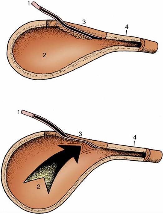
FIG. 5.31 Top, The ureterovesical junction. Bottom, Because of its oblique passage through the wall, the ureter is compressed (arrow) as the intravesical pressure rises. 1, Ureter; 2, bladder lumen; 3, bladder wall; 4, bladder neck.
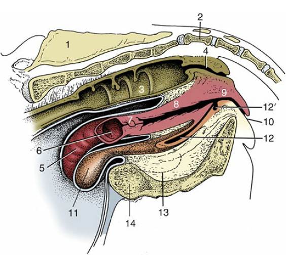
FIG. 5.32 Median section of the bovine pelvis.
1, Sacrum; 2, first caudal vertebra; 3, interior of rectum; 4, anal canal; 5, exterior of right uterine horn; 6, interior of stump of left uterine horn; 7, cervix; 8, vagina; 9, vestibule; 10, vulva; 11, exterior of bladder; 12, urethra; i2', suburethral diverticulum; 13, obturatorforamen; 14, pelvis symphysis.
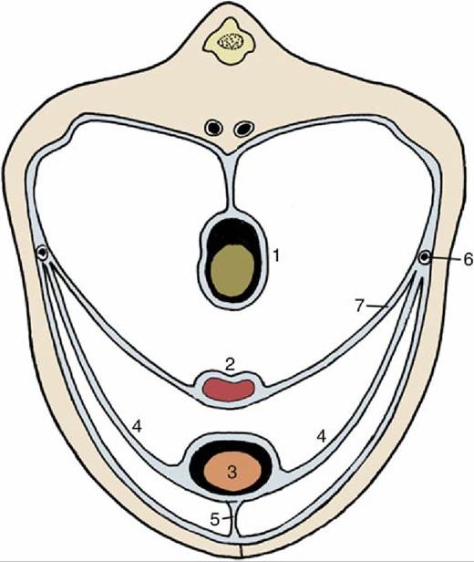
FIG. 5.33 Peritoneal disposition in the caudal part of the abdomen. 1, Colon; 2, uterus; 3, bladder; 4, lateral vesical ligaments; 5, median vesical ligament; 6, ureter; 7, broad ligament of uterus (mesometrium).
The bladder muscle is arranged in three sheets that exchange fascicles. The muscle is probably entirely detrusor—available to squeeze and empty the bladder—and fails to form an often described internal sphincter. Many writers now believe that, in place of this sphincter, some muscle bundles form a series of arcades whose summits are directed toward the orifice; they therefore dilate rather than occlude the exit when they contract. If this latter belief is accurate, continence depends on the tension passively exerted by the elastic elements within the mucosa and on the action of the external sphincter, the striated urethralis. This interpretation is consistent with the demonstration that in certain species (dog, goat) the proximal part of the urethra forms part of the urine reservoir, expanding as the bladder fills. The functional boundary between bladder and urethra would thus appear to be represented by the cranial limit of the urethralis in these species.
Autonomic fibers reach the bladder through the sympathetic hypogastric and parasympathetic pelvic nerves; the latter innervate the detrusor muscle. Sensory fibers are routed through the pudendal nerve. The main blood supply is from the vaginal (or prostatic) artery, but, as has been mentioned, it is supplemented by the reduced umbilical arteries.
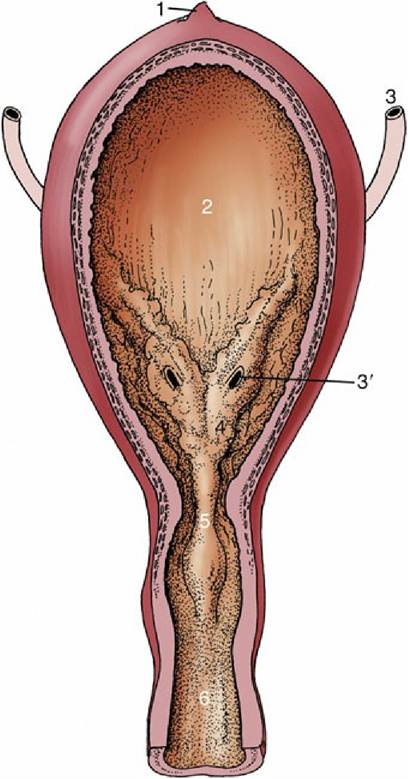
FIG.
5.34 The interior of the urinary bladder. 1, Scar of urachus; 2, bladder; 3, ureter; 3,, ureteric orifice;4, trigone of bladder; 5, urethral crest; 6, urethra.
The Female Urethra
The female urethra runs caudally on the pelvic floor below the reproductive tract. It passes obliquely through the vaginal wall to open ventrally at the junction of vagina and vestibule (Fig. 5.35). It is conspicuously short and wide in mares. In some animals, such as the cow and sow, it opens together with a suburethral diverticulum (Fig. 5.32/12'), and in others, such as the bitch, it opens on a hummock. Both arrangements create difficulties when catheterization of the bladder is attempted.
When a diverticulum is present, it is enclosed within the urethralis, which surrounds the urethra along most of its length. The cranial fascicles of the urethralis encircle the urethra, while the caudal ones support it within U-shaped loops that arise and end on the vaginal wall. Contraction of this part of the muscle closes the urethra by pressing the two organs together and also narrows the vagina. The urethralis obtains a somatic innervation through the pudendal nerve, but sympathetic and parasympathetic involvement is also described. The urethral submucosa contains many veins that constitute a form of erectile tissue that may contribute to continence by assisting mucosal apposition. These features apart, the structure of the urethra continues that of the bladder.