THE RUMINANT STOMACH
1. How do the camel and llama stomachs differ from those of sheep and cattle?
2. What kind of epithelium lines the forestomachs of the ruminant?
3. Which side of the cow would be used to detect rumen motility or observe rumen distention from tympanites?
4.
Why is bloat a hazard to breathing?5. Which stomach compartment is largest in newborn calves? In adult ruminants?
6. At what age do calves begin ruminating (assuming they have access to roughage)?
7. Where are boluses first deposited when they enter the rumen? Why are ingested, pointed, hardware items a hazard?
8. What is the function of the reticular groove?
9. How many contractions of the rumen ought to be observed each minute? Where can the contractions be felt?
0. What was the name of the Jersey steer that made the rumen fistula famous?
11. What are functions of the ruminant stomach compartments?
Animals that regurgitate and remasticate their food are called ruminants. There are two suborders of ruminant animals: (1) Ruminantia, which includes the deer, moose, elk, reindeer, caribou, antelope, giraffe, musk ox, bison, cattle, sheep, and goat and (2) Tylopoda, which includes the camel, llama, alpaca, and vicuna. The principal difference between the two suborders is that Tylopoda do not have an omasum. Another difference is that Tylopoda have areas of cardiac glands that open into ventral sacculated surfaces of the reticulum and rumen. These small sacs have given rise to the myth that the camel stores water in its rumen, but no evidence has been found to support the idea that more water is present in the camel rumen than in the rumen of other ruminants.
The ruminant stomach is adapted for fermentation of ingested food by bacterial and protozoan microorganisms. Energy is obtained through fermentation that would not otherwise be made available. In their natural environment, the diet of ruminants includes mostly growing, mature, or dried grass, and the mammalian digestive enzymes cannot digest the cellulose in these materials.
Microbial enzymes, however, can digest the plant cells through the fermentation process. Fermentation requires controlled conditions for a maximum rate of degradation; these are provided through appropriate secretions, motility, and body temperature. Regurgitation and remastication (associated with rumination) assist fermentation by providing more finely divided material and thus a greater surface area for microbial digestion. Foraging ruminants often seize and swallow food over a prolonged period, with only a relatively short time given to mastication. Remastication is done during times of relative quiescence. Resalivation is also accomplished during remastication, and the additional saliva is also beneficial to the fermentation process.Structure and Function
The ruminant stomach is composed of four compartments: (1) rumen (paunch), (2) reticulum (honeycomb), (3) omasum (many plies), and (4) abomasum (true stomach). The relationship of the compartments to each other is shown in Figures 12-37 and 12-38. The first three compartments are also known as the forestomach, because they precede the so-called true stomach. The forestomach is lined with stratified squamous epithelium and constitutes the nonglandular region of the stomach (see Figure 12-7). The rumen occupies a prominent portion of the viscera on the left side of the animal; its relationship to the thoracic viscera is shown in Figure 12-37. Note the proximity of the reticulum to the heart. The viscera, as seen from the right side, is shown in Figure 12-38. The abomasum is mostly on the right side. Tripe, a food product, is made from the rumen and reticulum after cattle are slaughtered. The rumen and reticulum are condemned for human food if they are ulcerated. The feeding of high-concentrate rations has been associated with ulceration and condemnation.
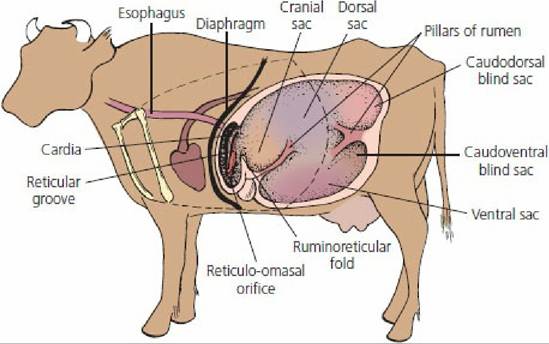
■ FIGURE 12-37 The stomach of cattle (left view).
The rumen and reticulum (shown) are two of the three compartments of the forestomach that precede the true stomach (abomasum). The reticulo-omasal orifice is the passageway to the third compartment known as the omasum. The rumen is divided into a number of sacs by muscular pillars. Pillar contraction is essential for movement of rumen content. The dashed line illustrates the extent of the rib cage.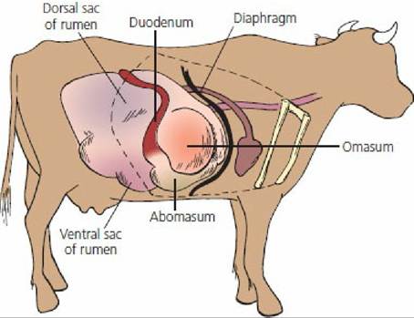
■ FIGURE 12-38 The stomach of cattle (right view). The omasum is the third compartment of the forestomach, which has a short omasal canal that connects the reticulo-omasal orifice with the omaso-abomasal orifice. The dashed line illustrates the extent of the rib cage.
The abomasum is the largest compartment of the newborn ruminant’s stomach (Figure 12-39). Forestomach development is associated with roughage intake and is lacking in calves that are fed only milk. Young ruminants usually begin ingesting limited roughage when they are 1 to 2 weeks old, and brief periods of rumination begin soon thereafter.
In the adult ruminant, the rumen is the largest forestomach compartment. It is separated from the much smaller reticulum by the ruminoreticular fold (see Figure 12-37). Food enters the rumen through the cardiac opening of the esophagus and is deposited in the cranial sac (atrium) of the rumen (see Figure 12-37). The next,contraction of the cranial sac transfers the contents into the reticulum, from where they can be “pumped” by contractions of the reticulum to: (1) the cardiac opening for regurgitation, (2) the omasum through the reticuloomasal orifice for transfer to the abomasum or for further digestion and absorption by the many plies of the omasum, or (3) more caudal parts of the rumen. Dense metal objects are often retained in the reticulum. If they are pointed objects, reticular contractions can result in their penetrating thoracic viscera (the heart or lungs), causing inflammation.
This condition is known as traumatic pericarditis (heart involvement) or, more commonly, hardware disease.The gastric groove (formerly called the esophageal groove) functions as a conduit for milk to bypass the nongalndular regions of the ruminant stomach and pass directly to the glandular abomasum. The gastric groove can be further subdivided into reticular, omasal, and abomasal grooves. The reticular groove (see Figure 12-37) transfers milk from the cardiac opening to the reticulo-omasal opening, from where it is conveyed to the abomasum through the omasal and abomasl grooves. Closure of the gastric groove is a reflex initiated when receptors in the mouth and pharynx are stimulated. The reflex loses its responsiveness with age. Although certain chemicals have been shown to bring about closure of the reticular groove in adult ruminants, no function has been described for it in the adult.
The various pillars of the rumen (see Figure 12-37) are muscular folds, which, when contracted, can move and mix large volumes of rumen content. One to two cycles of rumen contraction occur each minute. These can be felt when the hand is placed into the left paralumbar fossa (depression cranial to the pelvic hooks or tuber coxae, caudal to the ribs and ventral to the lumbar vertebrae). Assessment of rumen function is often made by this technique. Recall that there is a reflex inhibition of gastrointestinal motility in the presence of peritonitis, gas distention, and pain.
A permanent opening into the rumen at the paralumbar location is known as a rumen fistula. These surgical interventions have been used for obtaining samples and for making various physiologic measurements for many years. The fistula openings are plugged during times of nonuse. A classic example of a rumen fistula is shown in Figure 12-40.

■ FIGURE 12-39 Relative sizes of the bovine stomach compartments at various ages.
A. Three days old. B. Four weeks old. C. Three months old. D. Adult. a, Rumen; b, reticulum; c, omasum; d, abomasum. (From Nickel,R, Schummer A, Seiferle E. The Viscera of the Domestic Mammals. 2nd edn. Berlin: Verlag Paul Parey, 1979.)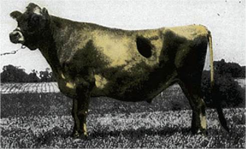
■ FIGURE 12-40 “ Bill,” a Jersey steer with a large rumen fistula. The animal was born in May 1942, the fistula was made in March 1943, and, after a leg injury, the steer was euthanized in January 1955. This photograph was taken in June 1954. When not in use, the fistula was kept closed with a pneumatic plug. (From Dukes HH. The Physiology of Domestic Animals. 7th edn. Ithaca, NY: Cornell University Press, 1955. Used by permission of the publisher, Cornell University Press.)
The functions of the ruminant stomach compartments can be summarized as follows:
1. The rumen allows for soaking and fermentation of bulk fibrous food and, because of its motility, the contents are continually mixed.
2. The reticulum serves as a pump that causes liquid to flow into and out of the rumen. The flow of liquid directs ingesta into the rumen, regulates its passage from the rumen to omasum, supplies moisture to rumen contents, and floods the cardia before regurgitation.
3. The omasum provides for continued fermentation and absorption (absorption enhanced by a large luminal surface related to the plies or leaves) and regulation of onward propulsion between the reticulum and abomasum.
4. The abomasum provides true stomach functions. Digestion of degraded roughages and concentrates begins for fermentation residues that have not been already absorbed. Also, the microbes of fermentation are prepared for their own digestion. Using microbes for nutrition of their host is an advantage ruminants have over nonruminant herbivores.
■