THE SALIVARY GLANDS
The parotid is clearly lobulated and has a firm texture and a yellow-gray or yellow-pink color. It is the largest salivary gland and extends ventrally from the base of the ear and wing of the atlas into the angle formed by the convergence of the maxillary and linguofacial veins and may possibly extend beyond because the maxillary vein frequently tunnels through the gland substance (Figure 18-7/14).
The cranial margin is largely contained by the caudal border of the mandible, but a thin flange extends some distance over the masseter directly ventral to the jaw joint, where it covers the parotid lymph nodes. The lateral surface is overlain by a well-developed fascia that gives attachment to the parotidoauricularis muscle. The deep surface is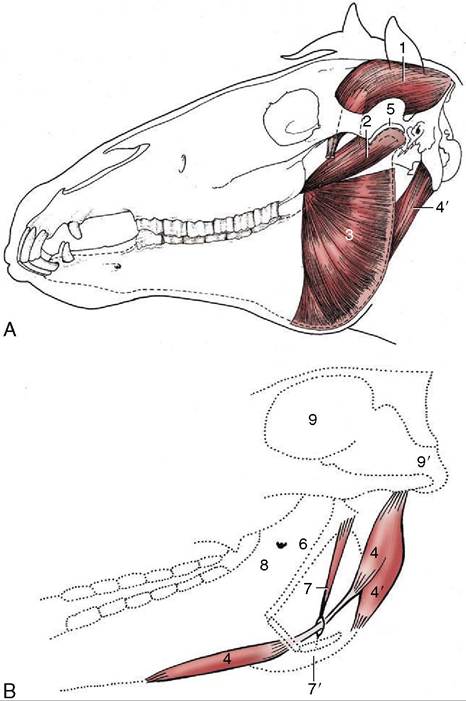
Figure 18-23 A, The deep masticatory muscles of the left side have been exposed by removal of the left mandibular ramus (stippled). B, Medial view of the right digastricus and some related structures. 1, Temporalis; 2, pterygoideus lateralis; 3, lateral surface of pterygoideus medialis; 4, digastricus; 4', occipitomandibularis; 5, left temporomandibular joint; 6, stylohyoid; 7, stylohyoideus; 7, insertion of 7 on thyrohyoid;
8, medial surface of right mandible and mandibular foramen;
9, cranial cavity; 9', foramen magnum.
related to the guttural pouch, the stylohyoid, the muscles that run to the corner of the jaw and open the mouth, and the combined insertion tendon of the brachiocephalicus and sternocephalicus, which separates it from the more deeply placed mandibular gland (see Figure 18-9).
The serous secretion of the parotid is drained by several sizable ducts that come together at the rostro- ventral angle of the gland to form a single channel. This crosses the tendon of the sternocephalicus before turning forward to run medial to the ventral border of the mandible. Accompanied by the facial vessels, it turns onto the face, where it ascends along the rostral margin of the masseter.
It first lies caudal to the artery and vein but later shifts rostral to them. It ends by opening into the vestibule opposite the third upper cheek tooth. The duct is relatively exposed in the last part of its course and may be damaged in superficial wounds. Leakage is most profuse when feeding stimulates the flow of saliva.The much smaller and crescentic mandibular gland extends from the basihyoid to the atlantal fossa and is thus partly under cover of the mandible (Figure 18-9/12 and Figure 18-30/5). The superficial relations include the parotid gland and the medial pterygoid, sternoce- phalic, digastric, and occipitomandibular muscles. Its deep location puts it out of reach on palpation. The mandibular duct is formed along the concave rostral margin of the gland by the confluence of several ductules. It runs rostrally, covered by the mylohyoideus, and follows the medial aspect of the sublingual gland until it opens on the floor of the mouth at the small sublingual caruncle. The secretion is mixed.
The sublingual gland lies directly below the oral mucosa, between the body of the tongue and the medial surface of the mandible, extending as a thin strip from the symphysis to the level of the fifth cheek tooth (Figure 18-30/1). It drains through numerous small ductules that open below the tongue.
Two rows of buccal glands are scattered along the dorsal and ventral margins of the buccinator. The glands of the dorsal series are more considerable and clump together caudally. Small salivary glands are found in the lips, soft palate, and tongue.