The Scrotum and Testes
The scrotum is perineal in position. The tail of the epididymis and the less salient associated pole of the testis point dorsocaudally by the anus and are readily palpable. The free border of the testis faces caudoventrally, and the attached border is closely applied to the surface of the thigh (Fig.
35.6).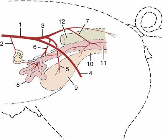
FIG. 35.5 The principal arteries supplying the left side of the female reproductive tract (schematic). 1, Aorta; 2, ovarian artery (a.) with cranial uterine branch; 3, internal iliac a.; 4, external iliac a. continued by femoral a. into left thigh; 5, umbilical a.; 6, left uterine a. crossing medial surface of external iliac a.; 7, vaginal a. with caudal uterine branch; 8, left uterine horn; 9, bladder; 10, urethra; 11, vagina; 12, rectum.
Castration: Male pigs are usually castrated within 2 weeks of their birth to prevent the development of the taint that characterizes the flesh of boars. It is now increasingly appreciated, in some countries at least, that the taint does not appear until after the usual age at slaughter and that castration is therefore pointless. Both the open and closed methods of castration are used with young pigs. In the former technique, which is preferred, the tunica vaginalis is incised, the ligament joining it to the epididymis divided, and the cord severed. This is the method employed with old boars. In the closed method (Fig. 35.7B), the scrotum is opened, the tunica vaginalis is left intact but freed from attachments, and the cord is transected close to the external inguinal opening. The situation of the scrotum explains the unusual length of the cord.
In pigs, descent of the testis commences about the 60th day of gestation, and regression of the extra-abdominal gubernaculum creates the conditions in which the testis is able to leave the inguinal canal by approximately the 90th day.
After a period of uncertainty, when the testis may move back and forth between the canal and the groin, a permanent position in the scrotum is adopted by full term. Abnormalities of gubernacular development and regression are common. Both excessive swelling and delayed regression may widen the canal abnormally, allowing a loop of intestine to slip into the vaginal cavity and thus creating an indirect inguinal or, should it reach so far, scrotal hernia. Surgical correction of this defect is generally combined with castration by the closed method. (The inguinal hernias occasionally seen in young gilts are associated with abnormal genital tracts that resemble those of bovine freemartins.)» TABLE 35.1
Guide to the Aging of Pig Fetuses
| Weeks Crown-Rump Length (cm) External Features | ||
| 2.5 | ≈1 | Limb buds forming |
| 4 | ≈2 | Tactile hair follicles appear; mammary primordium present |
| 5 | ≈3.5 | Palate fused; facial clefts closed |
| 6 | ≈6.5 | Prepuce and scrotum, or labia and clitoris present |
| 7 | ≈9 | Eyelids fused; intestines returned to abdomen |
| 13 | ≈24 | Eyelids separated |
| Full term | On average 114 days | |
From Evans HE, Sack WO: Prenatal development of domestic and laboratory animals. Growth curves, external features, and selected references. Anat Histol Embryol 2:11-45, 1973.
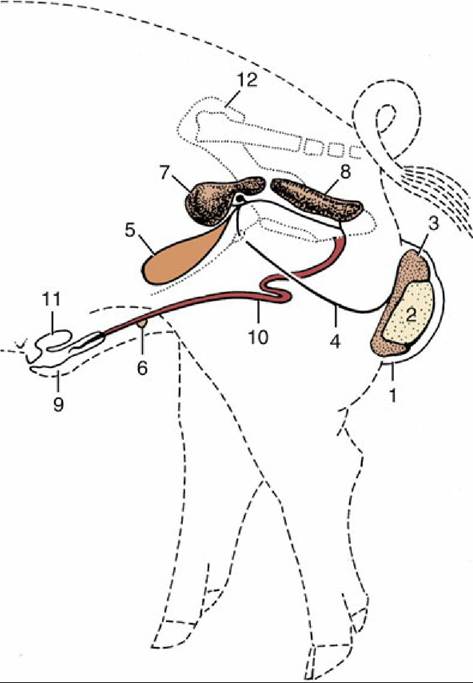
FIG. 35.6 Reproductive organs of the boar (schematic). 1, Scrotum; 2, left testis; 3, tail of epididymis; 4, deferent duct; 5, bladder; 6, rudimentary teat; 7, vesicular gland covering the small body of the prostate; 8, bulbourethral gland; 9, prepuce; 10, penis; 11, preputial diverticulum; 12, right hip bone.
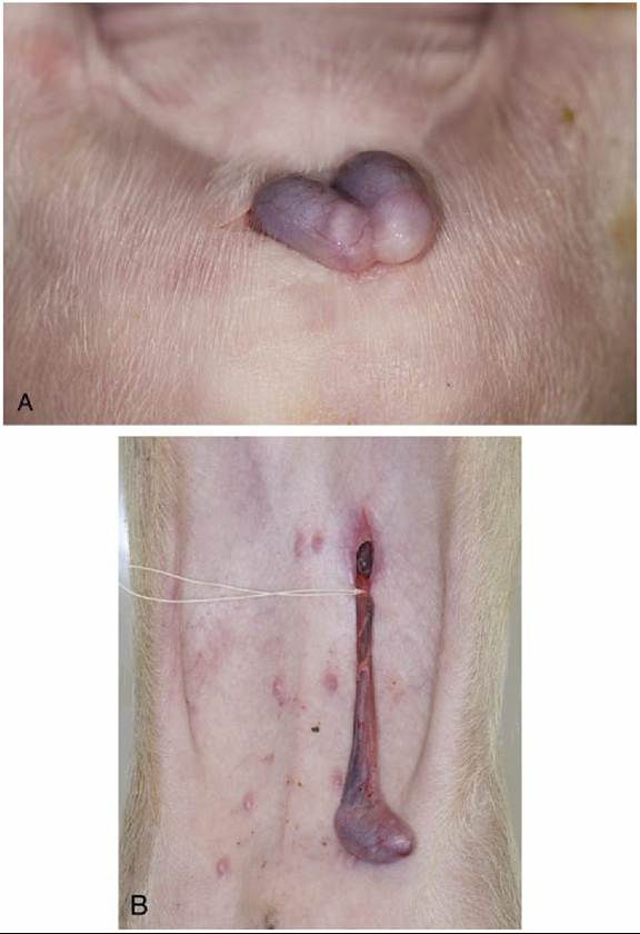
FIG. 35.7 (A) Open castration method of a newborn piglet. (Note: The parietal layer of vaginal tunic is still intact.) (B) The closed castration method in a 5-week-old piglet (also performed in case of an inguinal hernia).