THE SEBACEOUS GLANDS
These produce a fatty secretion (sebum) that lubricates and waterproofs the skin and coat. It also promotes the spread of sweat, retards bacterial growth, and, in certain instances, serves as a territorial marker that is recognized by other members of the species.
The odor of the wet dog is due to these glands. Certain substances (pheromones) present in sebum are known to be sexually attractive; the rate of production is controlled by steroid hormones (androgens generally promote secretion, and estrogens retard secretion). A good illustration of a selective effect of androgens is found in the reaction of the so-called acne region of the human adolescent.The sebum of the fleece of sheep is collected and processed; known as lanolin commercially, it is used as a base for ointments, in cosmetics, and as a cleansing agent in soaps. The secretions of certain specialized glands (e.g., the preputial glands of musk deer and the
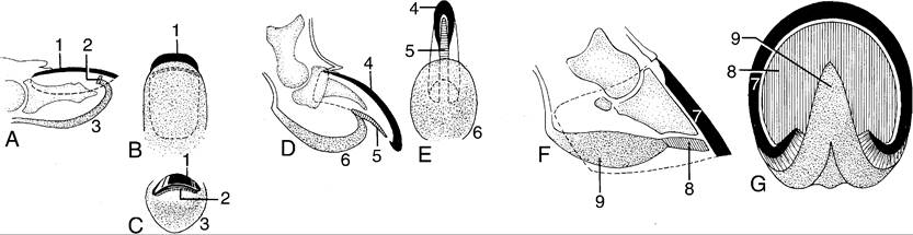
Figure 10-18 Schematic representation of nail, claw, and hoof. A-C, Longitudinal section, palmar surface, and head-on view of human fingertip. D, E, Longitudinal section and palmar surface of canine claw. F, G, Longitudinal section and ground surface of equine hoof. 1, Nail (wall); 2, "sole horn" of nail; 3, bulb of finger; 4, wall of claw; 5, "sole" of claw; 6, digital pad; 7, wall of hoof; 8, sole of hoof; 9, frog.
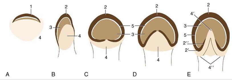
Figure 10-19 An interpretation of the phylogenetic "development" of the horn structures associated with the distal phalanx. A, A human fingertip. B, Pig. C, Rhinoceros. D, Tapir. E, Horse. 1, Nail; 2, wall of hoof; 2', 2", heel and bar (of horse); 3, sole; 4 footpad (bulb in human finger and pig); 4, 4", frog and bulbs of the heels (of horse); 5, white line.
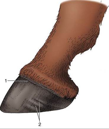
Figure 10-20 Equine hoof.
1, Periople; 2, rings indicating uneven horn growth.anal glands of the civet) have long been collected for use by the perfume industry.
The major localized accumulations of sebaceous glands that are of a size visible to the naked eye found in domestic animals are listed; several are associated with skin pouches.
Circumoral Glands (Figure 10-11)
These large glands are found in the lips of cats, which use them to mark their territories. The secretion is deposited directly by the animal rubbing its head against an object or ingratiatingly against its owner and indirectly after transference to the body during grooming.
Horn Glands (Figure 10-23)
These musk or scent glands are present in goats of both sexes, caudomedial to the horn base (or at the corresponding site in polled animals). They are larger and more productive in the breeding season; stimulated by testosterone, those of males produce a secretion with an
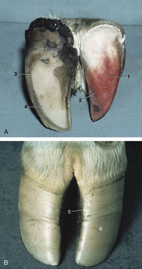
Figure 10-21 A, Bovine foot, palmer view. B, Bovine foot, dorsal view. The horn shoe (epidermis) has been pulled off one digit, exposing the dermis. 1, Dermis of bulb; 2, dermis of sole; 3, horn of bulb; 4, horn of sole; 5, dorsal border of hoof; 6, abaxial surface of hoof.
odor so pungent that some owners insist on their surgical removal.
Glands of the Infraorbital Pouch (Figure 10-24)
These glands are contained in a cutaneous pouch rostral to the eye and opening Ventrolaterally on the face of sheep. The pouch wall contains both sebaceous and tubular serous glands whose mixed secretion stains the skin when it escapes from the pouch. The glands, which serve as territorial markers, are larger in rams.
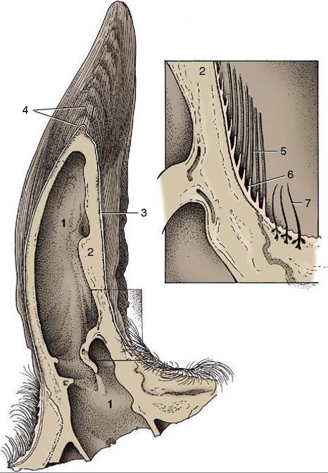
Figure 10-22 Longitudinal section of bovine horn. 1, Caudal frontal sinus extending into horn; 2, cornual process of frontal bone; 3, combined periosteum, dermis, and noncornified stratum of epidermis; 4, horn tubules separated by intertubular horn; 5, horn tubules (inset); 6, dermal papilla; 7, hair.
Carpal Glands (Figure 10-25)
These are present in pigs and cats.
In pigs they surround several cutaneous invaginations on the mediopalmar aspect of the carpus. They are found in both sexes and serve to indicate territorial claims; boars are said to make particular use of them when “marking” sows during copulation.The location of the glands in cats is marked by a tuft of a few tactile hairs proximal to the carpal pad. The site is betrayed by a palpable thickening of the skin (Figure 10-15, B/5).
Glands of the Interdigital Pouch (Figure 10-26)
Interdigital pouches are found on the forelimbs and hindlimbs of sheep of both sexes. The pouches are tubular invaginations of the skin whose walls contain branched sebaceous and serous glands. The waxy secretion is discharged through a single opening above the
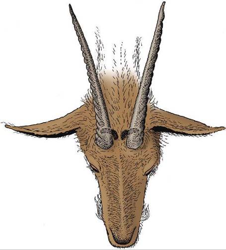
Figure 10-23 Horn glands caudomedial to the base of the horns in the goat.
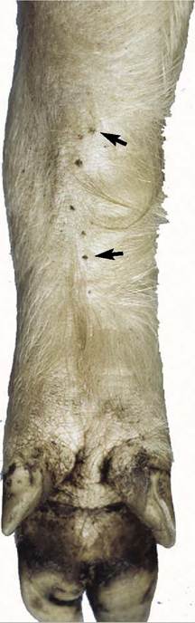
Figure 10-25 Carpal glands (arrows) of the pig, palmar view.
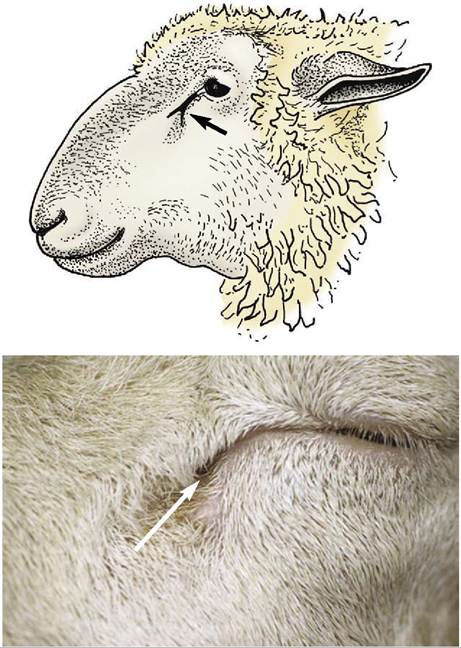
Figure 10-24 Infraorbital pouch (arrow) of the sheep.
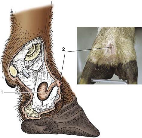
Figure 10-26 Interdigital pouch (1) of the sheep and its opening (2).
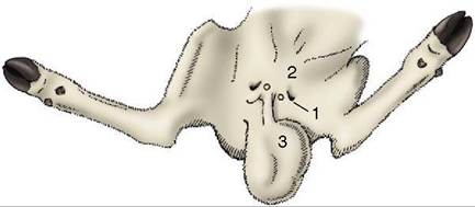
Figure 10-27 Inguinal region of the ram. 1, Inguinal pouch;
2, rudimentary teat; 3, scrotum.
id="Picutre 497" class="lazyload" data-src="/files/uch_group31/uch_pgroup304/uch_uch7232/image/image487.jpg">
Figure 10-28 Location of the tail glands of the dog.
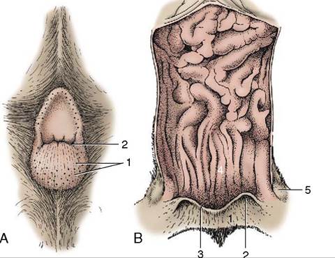
Figure 10-29 A, Cutaneous zone of the canine anal canal. B, Feline anal canal opened dorsally. 1, Cutaneous zone with circumanal glands forming a ring around the anus of the dog; 2, opening of the right anal sac; 3, anocutaneous line; 4, columnar zone; 5, right anal sac.
hoofs and serves as a “trail marker.” Many gregarious wild species have similar glands.
Glands of the Inguinal Pouch (Figure 10-27)
Inguinal pouches, found near the base of the udder or scrotum of sheep, contain both sebaceous and sweat glands. The secretion escapes as a brown waxy substance whose odor may assist the lamb to find the udder.
Preputial Glands (Figure 35-11)
Sebaceous and apocrine sweat glands within the prepuce produce secretions that combine with desquamated cells to form the crumbly substance known as smegma. They are best developed in the boar, in which they are massed within a dorsal diverticulum of the preputial cavity (see Figure 35-11/5). Their secretion gives the boar its characteristic odor. They are present but less offensive in other species (which lack the diverticulum).
Tail Glands (Figure 10-28)
Collections of large sebaceous and serous glands are found in an oval patch on the dorsal surface of the tail of certain carnivores. The skin over these glands is often defined by its sparser hair and yellowish color. Activity is greatest during the breeding season. The patch is situated more proximally in cats, toward the root of the tail, than in dogs (Figure 10-28).
Circumanal Glands (Figure 10-29)
These sebaceous glands are restricted to the perianal skin of certain carnivores, including dogs, where they drain into (and are believed to influence) special sweat glands. It is probably their secretion that excites the particular attention paid to the anal region when dogs confer. It has been suggested that some of these glands have an endocrine function.
Glands of the Anal Sacs (Figure 10-30)
Sebaceous and serous glands are found in the walls of the anal sacs, cutaneous pouches that open beside the anus of carnivores (Figure 10-29/2). The secretion, which is particularly foul-smelling, is expressed during defecation and apparently serves as a marker. It is well- known that skunks can forcefully expel the contents of the sacs to fend off aggressors.