The skeletal basis of the elbow joint is provided by the distal end of the humerus and proximal parts of the radius and ulna (Fig. 23.6A).
Both epicondyles of the humerus may be palpated without much difficulty, but the medial one is especially prominent and projects to the inner aspect of the olecranon. The condyle, identifiable more distally, presents a deep fossa for the anconeal process of the olecranon (Fig.
23.9/4 and 6). A shallow radial fossa occupies the corresponding site on the cranial aspect.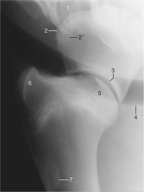
FIG. 23.7 Lateral radiograph of a shoulder joint. 1, Sixth cervical vertebra; 2, supraglenoid tubercle of scapula; 2', coracoid process; 3, glenoid cavity; 4, trachea; 5, head of humerus; 6, superimposed greater, lesser, and intertubercular tubercles; 7, deltoid tuberosity.
The powerful olecranon of the ulna rises high above the joint to project on the lower part of the fifth rib (or following space) and is therefore a less direct guide to the position of the articulation. The much reduced shaft of the ulna tapers distally to fuse and ultimately submerge with the shaft of the radius, but it leaves open an interosseous space in the proximal forearm. The proximal extremity of the radius is expanded and engages with the cylindrical humeral condyle. It also has medial and lateral eminences that furnish attachment to the collateral ligaments. The radial tuberosity is present to the front (Fig. 23.9/8). Both collateral ligaments may be palpated, although the medial one is covered by the relatively thick pectoralis transversus. A cranial division of this ligament represents a vestige of the pronator teres.
The shape of the articular surfaces and the presence of stout collateral ligaments restrict movement of the elbow joint to flexion and extension in a sagittal plane. The equine elbow is a good example of the "snap" joint, which abruptly moves from a stable to a more mobile position. This character depends on two features of its construction. The first is the unequal curvature of the humeral surface such that the radius of curvature of the central part is longer than those of the parts in front and behind, which are in contact with the radius in the more flexed and more extended positions of the joint. The second is that the collateral ligaments insert eccentrically on the humerus and are taut only in the intermediate position (Fig. 23.10).
The joint is most conveniently punctured by passing a needle between the lateral epicondyle and the olecranon into a caudal pouching of the joint capsule within the olecranon fossa.
The muscles of the arm that operate the elbow joint are arranged in flexor and extensor groups.