The Skeleton and Joints
The skeleton comprises the metacarpal bones and the proximal, middle, and distal phalanges. The metacarpophalangeal and the proximal and distal interphalangeal joints linking these bones are commonly referred to as the fetlock, pastern, and coffin joints.
A pair of proximal sesamoid bones enlarges the concavity of the fetlock joint, and a single distal sesamoid bone enlarges that of the coffin joint.The metacarpal skeleton comprises second, third, and fourth metacarpal bones. The third bone, the cannon bone, is much stronger than the other two and is the functional element. It carries a prominent tuberosity on its dorsal surface just distal to the joint. The bones to each side, generally known as the splint bones, are much reduced in size. Each has a small proximal base that continues into a tapering shaft. In young animals the splint and cannon bones are joined by fibrous tissue; this generally later ossifies, and the upper parts of the shafts are then fused together. The process is often accompanied by an acute inflammation (a condition known as "splints"), which leaves a palpable—and often visible—blemish on the dorsal surface.
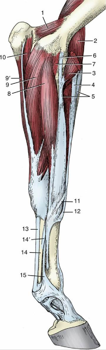
FIG. 23.18 Distal muscles of the left forelimb, medial view. 1, Anconeus; 2, brachialis; 3, biceps; 4, lacertus fibrosus; 5, extensor carpi radialis; 6, long part of medial collateral ligament (pronator teres); 7, short part of medial collateral ligament; 8, flexor carpi radialis; 9 and 9', humeral and ulnar heads of flexor carpi ulnaris, respectively; 10, ulnar head of deep digital flexor; 11, tendon of extensor carpi obliquus; 12, tendon of extensor carpi radialis; 13, tendon of superficial digital flexor; 14, tendon of deep digital flexor; 14', accessory (check) ligament; 15, interosseus.
The tapering second and fourth metacarpals end in slight but easily palpable buttons three- quarters of the way down the cannon (see Fig.
2.49B). The lower parts of their shafts are free, andwhen a break occurs, it is a simple matter to remove the fragment below the fracture line.
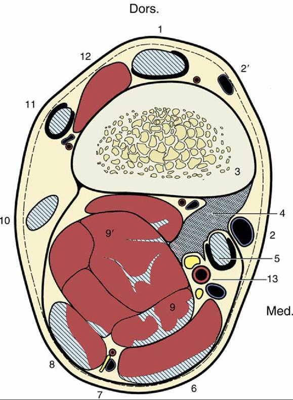
FIG. 23.19 Transverse section of the right forearm 6 cm proximal to the proximal border of the accessory carpal, to demonstrate the topography of the accessory (check) ligament of the superficial digital flexor; looking distally. The hatched blue areas are tendons or tendinous tissue, and the dark pink areas are muscle tissue. 1, Extensor carpi radialis; 2 and 2', cephalic and accessory cephalic veins, respectively; 3, radius; 4, accessory (check) ligament of superficial digital flexor; 5, flexor carpi radialis; 6, flexor carpi ulnaris; 7, ulnar nerve and collateral ulnar vessels; 8, ulnaris lateralis; 9 and 9', superficial and deep digital flexors, respectively; 10, 11, and 12, lateral, common, and oblique extensors, respectively; 13, median artery, medial and lateral palmar nerves; Dors., dorsal; Med., medial.
The third metacarpal bone is exceptionally robust. It is oval in cross section (which distinguishes it from the longer but more rounded cannon bone of the hindlimb), and its thick compacta attests to its tremendous strength; it is in fact one of the strongest elements of the skeleton (see Fig. 23.46/1). Despite the obvious strength of the cannon bone, longitudinal fractures of the distal extremity are common racing injuries, more often involving the lateral than the medial side and the forelimb rather than the hindlimb. The degree of involvement of the joint surface is an important factor in prognosis.
The distal extremity presents an axially keeled condyle that articulates with the proximal phalanx and the paired sesamoid bones. When viewed from the side, the condyle encompasses some 220 degrees of a circle, which is evidence of the great range of flexion and extension—the only movements allowed. The articular surface to each side of the keel is interrupted by a slight ridge that separates the more strongly curved palmar area from the larger dorsal one.
» TABLE 23.6
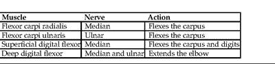
Muscles of Forearm: Flexors
The proximal sesamoid bones are three-sided pyramids whose bases face distally (Fig. 23.20/10). The dorsal (articular) surface of each lies against the condyle, the palmar (flexor) surface tilts axially and faces the flexor tendons that ride over it, and the abaxial surface is hollowed for the reception of the thick branch of the interosseous (see further on). The palmar aspects of the bones are converted by thick fibrous tissue (palmar ligament) into a single bearing surface over which the flexor tendons change direction. Although close to the proximal phalanx, the sesamoid bones do not articulate with it.
The proximal sesamoids fracture, commonly at the apical surface, most often of all the bones in the forelimb, followed in frequency by the metacarpal and carpal bones. These fractures are known in racetrack practice as "the big three" for which, when serious, horses pay with their lives. Fractures of proximal sesamoids are largely because of excessive pressure from the deep digital flexor tendon.
The strong proximal phalanx (also called PI) is compressed from front to back and is wider proximally than distally. Its proximal extremity is hollowed and deepened axially by a groove that allows it to conform to the condyle of the large metacarpal bone. Palpable tubercles to each side receive the collateral ligaments of the fetlock joint. The distal end is shaped as two condyles separated by a shallow axial groove and presents similar but smaller tubercles for the collateral ligaments of the pastern joint. The palmar surface of the bone is roughened for the attachment of several ligaments; a large triangular area and various smaller ones to each side stand out (Fig. 23.20B/11, 11', 11", and 11'").
The middle phalanx (PII) is generally similar to PI but, being only half as long, is proportionately very robust.
Both extremities are of equal width. The proximal articular surface—hollowed with a slight axial ridge—is the reciprocal of the lower end of PI, whereas the distal one—two condyles separated by a groove—mimics that of PI. The distal articular surface extends onto the palmar aspect, where it articulates with the distal sesamoid bone. There are proximal collateral tubercles on PII for the collateral ligaments of the pastern joint; the corresponding distal sites from which the collateral ligaments of the coffin joint arise are excavated. The proximopalmar border presents a smooth area (Fig. 23.20/12 ") that is enlarged in the natural state by a complementary fibrocartilage that forms a bearing surface for the deep flexor tendon (see further on). The fibrocartilage enlarges the articular surface of the pastern joint and gives attachment to several ligaments. Fractures of the middle phalanx are seen more in Quarter horses and more commonly in the hindlimb.The distal phalanx (PIII, coffin bone) generally conforms to the interior of the hoof in which it resides, "as in a coffin." It is wedge shaped: sharp distally and to the sides and blunt proximally and toward the back. The dorsal (parietal) surface is convex from side to side and lies against the dermis that unites it to the inner surface of the hoof wall. It tapers caudally into medial and lateral palmar processes that are notched (or perforated) and grooved for the dorsal terminal branches of the digital arteries and accompanying nerves (Fig. 23.20/13"). Depressions for the collateral ligaments of the coffin joint are present proximodorsal to the processes. The palmar (sole) surface is slightly concave to fit the domed sole of the hoof. Both parietal and sole surfaces are very porous to allow the passage of numerous small arteries from the interior of the bone into the overlying dermis. The articular surface, consisting of two fossae separated by an axial ridge, faces proximally. Its dorsal border tapers to an extensor process, the highest point of the bone, where the common digital extensor tendon is attached.
The palmar border is extended by a narrow articular zone for the distal sesamoid bone, which, in contrast to the proximal sesamoids, articulates with both middle and distal phalanges. Just distal to this, two prominent foramina lead to a U-shaped canal within the bone that contains the anastomosis of the terminal palmar branches of the digital arteries. The deep flexor tendon ends on the semilunar crest just distal to the foramina (Fig. 23.20/15).The flat cartilages (of the hoof), which surmount and continue the palmar processes, lie mainly against the inner wall of the hoof, but their proximal borders are free, subcutaneous, and palpable to each side of the pastern joint (Fig. 23.20B/14).
The distal sesamoid (navicular) bone (Fig. 23.21/3) is boat shaped with straight proximal and convex distal borders. Its dorsal (articular) surface contacts the distal end of PII; a narrow distal facet touches PIII. The palmar (flexor) surface faces the wide tendon of the deep flexor, providing it with yet another bearing surface as it bends toward its attachment at the semilunar crest on the undersurface of PIII. The navicular bone enlarges the distal articular surface of the coffin joint (see Fig. 23.24/7' and 7").
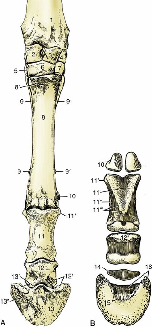
FIG. 23.20 Skeleton of the distal part of the forelimb. (A) Left limb, dorsal view. (B) Palmar view. 1, Radius; 2, radial carpal; 3, intermediate carpal; 4, ulnar carpal; 5, 6, and 7, second, third, and fourth carpals, respectively; 8, large metacarpal bone; 8', metacarpal tuberosity; 9 and 9', medial and lateral splint bones, respectively; 10, proximal sesamoid bones; 11, proximal phalanx; 11', proximal tubercle; 11", attachment of distal digital annular and abaxial palmar ligaments; 11'", attachment of axial palmar and oblique sesamoidean ligaments; 12, middle phalanx; 12', attachments of collateral ligament of coffin joint; 12", bearing surface for deep flexor tendon; 13, distal phalanx; 13', extensor process; 13", parietal groove; 14, navicular bone; 15, sole foramen and semilunar crest for attachment of deep flexor tendon; 16, palmar process and attachment of distal navicular ligament.
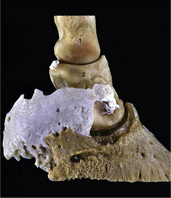
FIG. 23.21 Hoof cartilage attached to palmar process of distal phalanx. 1, 2, and 3, Proximal, middle, and distal phalanges, respectively; 4, hoof cartilage.
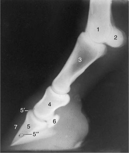
FIG. 23.22 Lateral radiograph of fetlock joint and digit. 1, Large metacarpal bone; 2, proximal sesamoid bones; 3, proximal phalanx; 4, middle phalanx; 5, distal phalanx; 5', extensor process; 5", canal containing terminal arterial arch; 6, navicular bone; 7, wall of hoof.
The fetlock joint is formed between the large metacarpal bone (PI) and the proximal sesamoid bones (Fig. 23.22). The large bones are connected by medial and lateral collateral ligaments, while additional smaller and triangular (collateral) ligaments anchor the sesamoid bones to the sides of the metacarpal condyle and the proximal tubercles of PI. A series of sesamoidean ligaments connects the bases of the sesamoid bones to the first phalanx and ensures that the sesamoids move against the metacarpal condyle in unison with PI. The series includes short and deepest ligaments to the proximopalmar border overlain by rather longer cruciate ligaments that end a little more distally, and these in turn are overlain by oblique ligaments that attach broadly to the central triangular area of the palmar surface of PI. Finally, an additional straight sesamoidean ligament, arising from the bases of the sesamoids, connects with the complementary fibrocartilage of PII (Fig. 23.23/4).
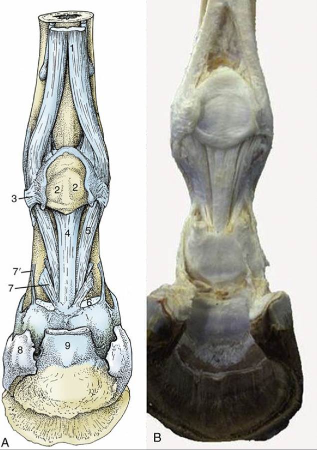
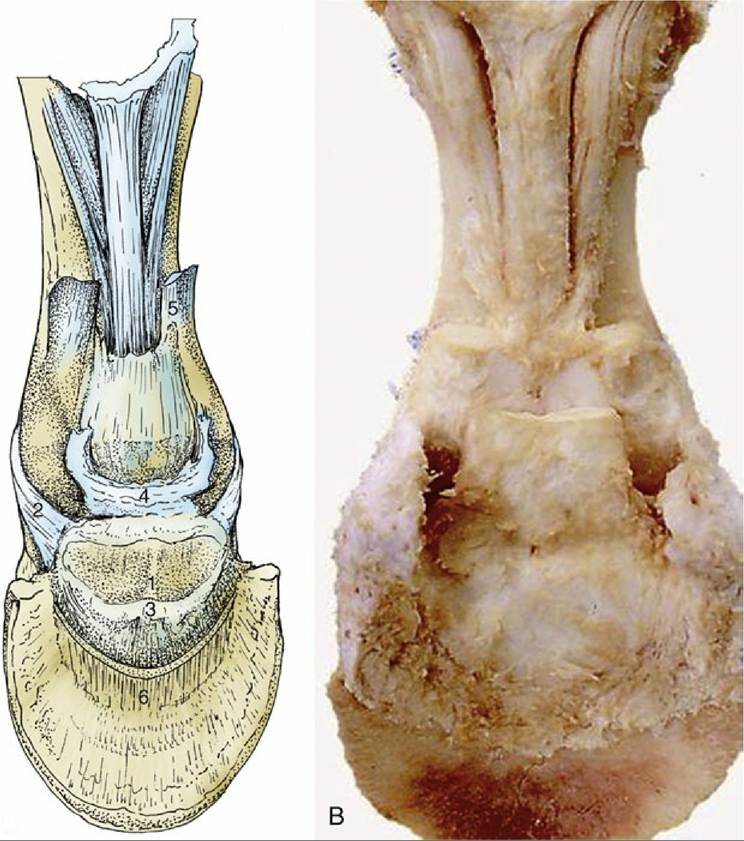
FIG. 23.23 (A) Structures supporting the fetlock joint (schematic). 1, Interosseus; 2, proximal sesamoid bones connected by thick palmar ligament; 3, collateral sesamoidean ligament; 4, straight sesamoidean ligament; 5, oblique sesamoidean ligament; 6, stump of superficial flexor; 7 and 7', axial and abaxial palmar ligaments of pastern joint, respectively; 8, hoof cartilage; 9, stump of deep flexor. (B) Real specimen.
The sesamoid bones are connected to each other by a thick palmar ligament that extends the bearing surface for the flexor tendons proximally by about 2 cm (Fig. 23.23/2). This extension supports the tendons when the sesamoids themselves slip below the condyle in maximal overextension of the fetlock joint (when the dorsal angle can be as small as 90 degrees). When the joint is fully flexed, the sesamoid bones lose contact with the condyle and ride up on the back of the metacarpal bone, where bone-to-bone contact is prevented by the proximal extension of the palmar ligament.
The joint capsule is capacious, and to allow for the fetlock's mobility, it extends large dorsal and palmar pouches proximally (see Fig. 23.26/7 and 7"). These lie against the shaft of the metacarpal bone and are easily punctured from the side; the end of the splint bone, the interosseus, and the sesamoid bone are convenient (almost visible) landmarks for entry into the palmar pouch. Another perhaps better place in the bowed limb is between the sesamoid bone and the metacarpus, directly through the collateral ligament of the flexed joint (see Fig. 23.27B (arrow) and C). Distentions of the joint known as wind puffs or galls manifest themselves at this site. The interior of the dorsal pouch contains a so-called capsular fold (see Fig. 23.26/7'). This arises from the shaft of the metacarpal bone and projects distally into the center of the pouch; its inflammation and enlargement can cause lameness. Short distal palmar pouches are palpable as small depressions in the angles between PI and the bases of the sesamoid bones.
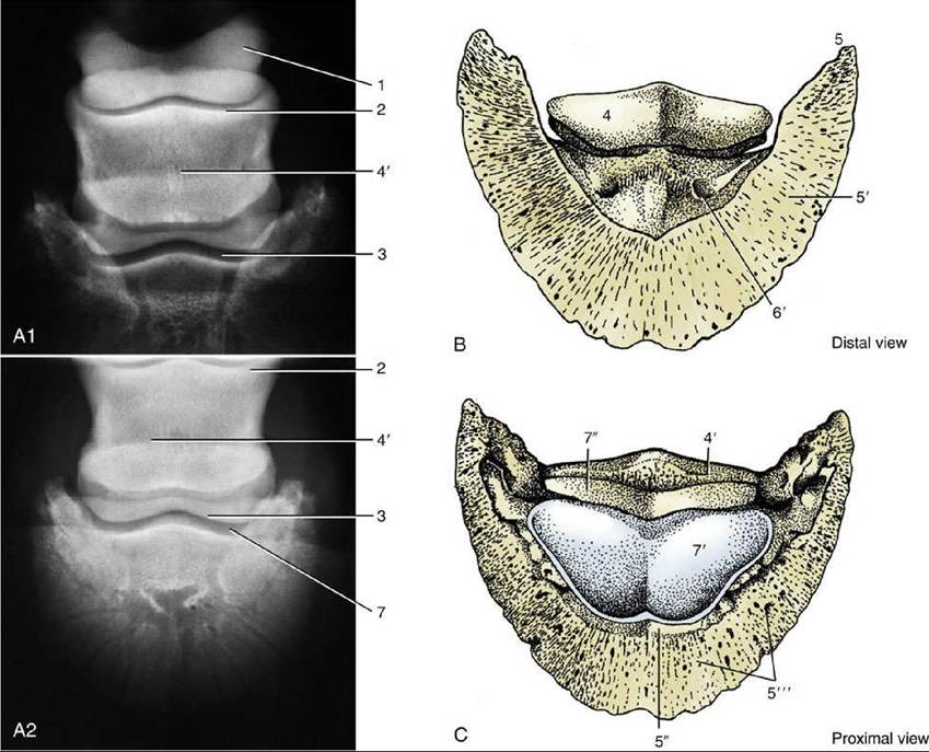
FIG. 23.24 (A1) and (A2) Dorsopalmar radiographs of hoof. (B) and (C) Palmar and dorsal surfaces of distal phalanx (PI) and navicular bone. 1, Proximal phalanx; 2, proximal contour of middle phalanx; 3, distal contour of middle phalanx; 4, navicular bone (its flexor surface in B); 4', proximal border of navicular bone; 5, palmar process of PIII; 5', palmar (sole) surface of PI; 5", extensor process and dorsal (parietal) surface of PI; 5'", dorsal surface; 6, sole foramen; 7, coffin joint; 7', articular surface of PI; 7", articular surface of navicular bone.
The movement of the pastern joint is much more restricted. Paired (axial and abaxial) palmar ligaments connect the palmar aspect of PI with the complementary fibrocartilage of PII (Fig. 23.23/7 and 7'); together with the straight sesamoidean ligament (Fig. 23.23/4) they limit overextension. The capsule is similar to that of the fetlock joint, but the pouches are smaller and only the dorsal one is accessible for puncture, again from the side. The radiographic appearance of the pastern and coffin joints is shown in Fig. 23.24(A1 and A2).
The coffin joint allows flexion and extension to about the same degree as the pastern joint. The collateral ligaments are short and thick and are solidly anchored at both ends to depressions in the bones. The navicular bone, an integral part of the joint, is suspended from the distal extremity of PI by the collateral navicular ligaments (Fig. 23.25/2) that cross the medial and lateral borders of PII and attach to the ends and proximal border of the navicular bone in a U-shaped fashion. A very short but wide distal navicular ligament (Fig. 23.25/3) connects the distal border of the navicular bone with PIII, attaching proximal to the prominent sole foramina. The capsule attaches to the articular margins of the three bones and resembles those of the other digital joints in having dorsal and palmar pouches. The pouches are small, and only the dorsal one is accessible for puncture (at the proximal border of the hoof); the procedure is not easy (Fig. 23.26C and D).
A
FIG. 23.25 (A) Ligaments of the navicular bone, palmar view (schematic). 1, Navicular bone; 2, collateral ligament of navicular bone; 3, distal navicular ligament; 4, connective tissue between coffin joint, digital sheath, and navicular bursa (see Fig. 23.26/15); 5, stump of superficial digital flexor; 6, stump of deep digital flexor. (B) Real specimen.
The incorporation of sesamoid bones in the fetlock and coffin joints divides the weight pressing onto the lower part of each joint over the phalanx and sesamoid bones. The elasticity of the sesamoid ligaments and the flexor tendons behind them allows the joint to yield slightly during foot impact. This is but one of several mechanisms designed to dissipate the concussion generated by so heavy and swift an animal. The concussive effects may be accentuated by poor conformation: upright pasterns and small feet (in relation to body size) are a combination encountered frequently in animals afflicted with navicular disease, a relatively common cause of lameness. This condition is characterized by erosion at the margins of the navicular bone, where its ligaments attach, and by inflammation and degeneration of the navicular bursa (Fig. 23.26/10) and the related part of the deep flexor tendon (Fig. 23.26/13). However, the exact pathogenesis is still debated, and different authorities give quite contradictory explanations.