The Skeleton of the Hindlimb
Pelvic Girdle
The pelvic girdle has been described with the trunk (p. 40) for the reason previously given.
Skeleton of the Free Appendage
The femur (os femoris; Fig. 2.58), the skeleton of the thigh, is the strongest of the long bones.
The proximal end curves medially so that the proximal articular surface, the head, is offset in relation to the long axis of the shaft. The femoral head is hemispherical and is joined to the shaft by a neck, which is best defined in the smaller species. The articular surface has a nonarticular area (fovea) to which the intracapsular ligament(s) attach(es); the fovea is round and central in the dog and is wedge-shaped and extended to the medial periphery in the horse. A large process, the greater trochanter (Fig. 2.58/3), is lateral to the head and rises level with the head in small animals but projects high above it in larger species (Fig. 2.58/3' and 3 "). The greater trochanter gives attachment to the bulk of the gluteal muscles, providing these extensors of the hip with a long lever arm. The trochanteric fossa lies between the trochanter and the femoral neck (Fig. 2.58/5) and the site of insertion of the small rotator muscles of the hip.The caudal aspect of the femoral shaft is flattened, but the other aspects combine in a continuous smooth surface. The borders between the flat and rounded areas are roughened for muscular attachments. Two processes mark the proximal half of the shaft. A low and rough lesser trochanter (Fig. 2.58/4) projects from the medial border and gives insertion to the iliopsoas muscle. The third trochanter located at the base of the greater trochanter (trochanter tertius; Fig. 2.58/4') is salient only in the horse and gives attachment to the gluteus superficialis. In the large animals the caudodistal part of the shaft has a deep supracondylar fossa that increases the area of origin of the superficial digital flexor (Fig.
2.58/7'). The same function is fulfilled by tuberosities in the dog.The distal extremity of the femur articulates with the tibia and the patella. The articulation with the tibia is accomplished by two condyles directed caudodistally and separated by a deep intercondylar fossa. The abaxial surfaces of the condyles are roughened and give attachment to the collateral ligaments of the stifle. The lateral condyle has a cranial depression called the extensor fossa (Fig. 2.58/12), which gives origin to the long digital extensor and peroneus tertius muscles, and a caudal depression (Fig. 2.58/13) for the popliteus. In the dog and cat the caudal aspect of each condyle is surmounted by a small flat facet for articulation with one of the small sesamoid bones (Fig. 2.58/11; formerly fabellae) in the origin of the gastrocnemius (see Fig. 17.3). A cranial trochlea (Fig. 2.58/6) articulates with the patella and extends proximally on the cranial surface. The bounding ridges are low and more or less equal in size in the dog and relatively larger and disparate in the horse and in cattle, in which the stouter medial ridge ends in a proximal enlargement (Fig. 2.58/6').
The patella, the kneecap, is a sesamoid developed within the insertion of the quadriceps femoris, the main extensor of the stifle. It is ovoid in the dog but prismatic in the horse and in cattle. The patella is extended medially and laterally by parapatellar cartilages in the fresh state.
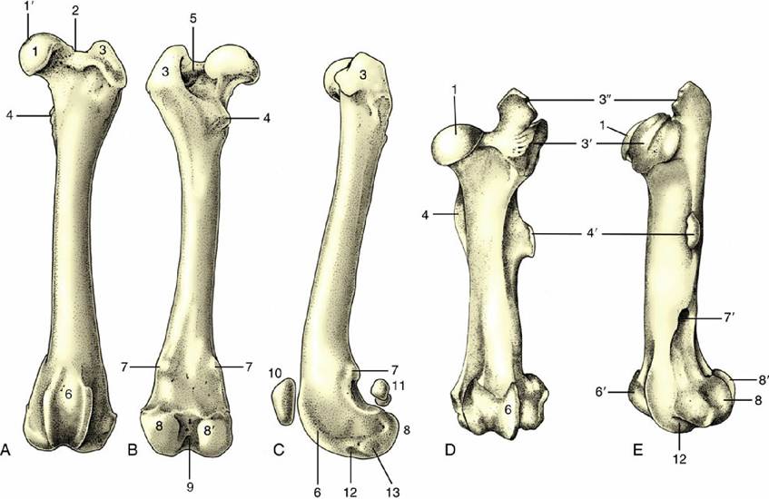
FIG. 2.58 Left femur of the dog. (A) Cranial, (B) caudal, and (C) lateral views. Left equine femur. (D) Cranial and (E) lateral views. 1, Head; 1', fovea; 2, neck; 3, greater trochanter; 3' and 3", cranial and caudal parts of greater trochanter; 4, lesser trochanter; 4', third trochanter; 5, trochanteric fossa; 6, trochlea; 6', enlarged proximal end of medial trochlear ridge; 7, supracondylar tuberosities; 7', supracondylar fossa; 8 and 8', lateral and medial condyles; 9, intercondylar fossa; 10, patella; 11, sesamoid bones (in gastrocnemius); 12, extensor fossa; 13, fossa for popliteus.
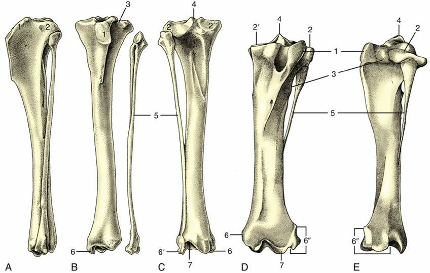
FIG.
2.59 Left tibia and fibula of the dog. (A) Lateral, (B) cranial, and (C) caudal views. Left equine tibia and fibula. (D) Cranial and (E) lateral views. 1, Tibial tuberosity; 2 and 2', lateral and medial condyles; 3, extensor groove; 4, intercondylar eminence; 5, fibula; 6 and 6', medial and lateral malleoli; 6", lateral malleolus in the horse (representing distal end of fibula); 7, cochlea.The skeleton of the leg consists of the tibia and fibula (Fig. 2.59), which, unlike the analogous elements of the forelimb, run side by side without any tendency to cross. The medial bone, the tibia, is always by far the larger of the two. The fibula is excluded from articulation with the femur and has only restricted contact with the hock skeleton.
The proximal extremity of the tibia presents two condyles divided by a caudal popliteal notch that has the popliteal muscle. Each condyle has a gently undulating articular surface for the corresponding condyle of the femur; a narrow intermediate nonarticular area carries a central eminence (Fig. 2.59/4). A depression of the eminence and less defined areas cranial and caudal to it indicate ligamentous attachments. The very robust tibial tuberosity (Fig. 2.59/1) on the cranial aspect of this extremity is a prominent landmark in life and is continued by a gradually subsiding crest. A groove (Fig. 2.59/3) lodging the tendons of certain muscles of the leg (crus) separates the tuberosity from the cranial aspect of the lateral condyle. Caudal to this groove, the edge of the condyle carries a small facet for articulation with the fibula, although in some species the joint space is obliterated by fusion.
The proximal part of the tibial shaft is three-sided, but more distally the bone is craniocaudally compressed. The change is brought about by the smooth surface that faces craniolaterally in its proximal part but then twists to face directly forward. The entire medial surface is subcutaneous and flat. The caudal surface is ridged for muscular attachment.
The distal extremity contains the cochlea (Fig. 2.59/7), which articulates with the trochlea of the talus. The central ridge and the flanking grooves of the cochlea have a craniolateral deflection, although the angle varies among species. A bony salience, the medial malleolus (Fig. 2.59/6), is present to the medial side of the cochlea. A similar lateral swelling found only in the horse represents the assimilated distal part of the fibula (Fig. 2.59/6"). In other species the corresponding feature (lateral malleolus) is provided by the fibula.
The fibula of the carnivores and the pig is reduced in robustness but not in length. It is separated from the tibia by an interosseous space that runs the whole length of the leg in the pig but is limited to the proximal half in the dog. The shaft of the fibula is reduced in the ruminants to a tear-shaped process fused to the lateral condyle of the tibia; the distal extremity is isolated as a small compact malleolar bone that forms an interlocking joint with the tibia, thus completing the articular surface for the talus. The flattened proximal head of the fibula of the horse is closely applied to the tibia, and the slender shaft that leads from it converges on the tibia but fades toward the middle of the
leg.
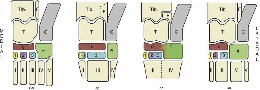
FIG. 2.60 The bones of the tarsal skeleton in the carnivores (Car), horse (eq), cattle (bo), and pig (su) schematic. Roman numerals identify the metatarsal bones, arabic numerals the distal tarsal bones. C, calcaneus; c, central tarsal bone; F, fibula; T, talus; Tib., tibia.
The tarsal bones are arranged in three tiers. The proximal tier consists of two relatively large bones: the talus medially and the calcaneus laterally. The middle tier comprises only a single central tarsal bone, but the distal tier consists of up to four bones, which are numbered in mediolateral sequence. The lateral fourth tarsal bone is constantly present and, being much deeper than the others, intrudes into the middle tier (Fig.
2.60).The talus (Fig. 2.61) has a proximal trochlear surface shaped to fit the tibia. The distal surface, which articulates with the central bone, is flattened in the horse and more rounded in other species. The calcaneus lies mainly lateral to the talus but extends a shelflike process (sustentaculum tali; Fig. 2.61/3') that overlaps the talus on its plantar surface and supports the deep digital flexor tendon. The larger part of the bone projects proximally behind the tibia as a free lever arm to which the common calcanean tendon attaches. The talus ends in a thickening that is the basis of the point of the hock (Fig. 2.61/3'') and corresponds to the human heel. The distal extremity of the calcaneus rests on the fourth tarsal bone (Fig. 2.61/6). The central tarsal bone is interposed between the talus proximally and the first, second, and third tarsal bones distally; its proximal surface conforms to the talus and is concave in most animals but flat in the horse. Its distal articular surface is flattened. The central and fourth tarsal bones fuse in ruminants.
The distal tarsal bones are not always separate: the first and second are fused in the horse, and the second and third are fused in ruminants. Individually irregular, these bones together form a more or less flattened disk interposed between the central tarsal and the metatarsal bones. The cuboidal fourth tarsal bone is interposed between the calcaneus and the lateral metatarsal bones; in some species it also gives support to the talus.
The remaining bones of the hindlimb closely resemble those of the forelimb. The metatarsal bones are longer (by about 20%) than the metacarpals and are more rounded in cross section. The first metatarsal is rudimentary in the dog, of which only a few breeds consistently possess a dewclaw in the hindlimb.