THE SMALL INTESTINE
The duodenum is short and closely attached to the abdominal roof by a short mesoduodenum. The initial portion continues from the pyloric part of the stomach and passes toward the right body wall before being deflected caudally to descend to a point between the right kidney and the pelvic inlet.
It then passes medially, behind the root of the mesentery, before ascending a short distance; it ends by bending ventrally to enter the mesentery, where it is continued as the jejunum. The more constant relations of the dog’s duodenum are to the liver at its origin, thereafter to the right body wall laterally, to the pancreas and later the right kidney medially, and, overall, to other parts of the intestinal mass. Although the first part of the duodenum is not expanded to form a distinct “duodenal bulb” or “cap” (so commonly the site of ulcers in people), its functional independence is retained.The jejunum and ileum are less closely fixed in position, but, although the arrangement of individual coils continually adjusts, this gut as a whole occupies a more or less constant position in the ventral part of the abdominal cavity (Figure 3-41). The coils are carried by the mesentery, which conveys the vessels and nerves; the mesentery is bunched at its root around the origin of the cranial mesenteric artery from the aorta and widens to the length of the gut at its other margin. The initial and final portions of the mesentery are shortest and ease the transitions with the relatively fixed duodenum at one end and with the ascending colon at the other (see Figure 3-40). The distinction between jejunum and ileum is arbitrary and perhaps unnecessary, for although certain progressive structural changes occur, these do not allow recognition of a sharp boundary. The convention that we follow limits the ileum to a short, relatively more muscular (and hence firmer) final portion with a direct peritoneal connection with the cecum.
Many anatomists from English-speaking countries assume a more or less equal division between the two parts.The jejunum fills those parts of the abdomen that are not preempted by other viscera. In the dog, in which the large intestine is relatively small, it lies more or less symmetrically about the midline, between the liver and stomach cranially and the urinary bladder caudally. It lies on the abdominal floor, though separated from the
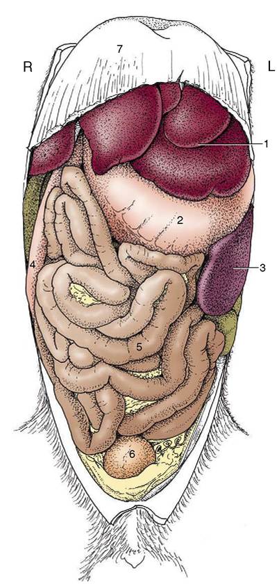
Figure 3-41 Ventral view of the abdominal organs of the dog after removal of the greater omentum. 1, Liver; 2, stomach; 3, spleen; 4, descending duodenum; 5, jejunum; 6, bladder; 7, diaphragm.
parietal peritoneum by the intervention of the greater omentum. The coils are quite mobile, and at first sight their disposition appears to be haphazard; closer inspection shows that there is some pattern to the arrangement. The mainly sagittal coils of the proximal part lie largely cranial to the more transverse coils of the distal part (see Figure 3-41). The ileum pursues a rather direct cranial, dorsal, and dextral course toward its junction with the large intestine. In life the intestine is not uniformly full, and at any moment most parts are flattened and molded by the pressures of adjacent viscera. The lumen may be locally obliterated, and when a passage is retained, it is more often than not reduced to a narrow channel along one margin: a “keyhole” form is seen
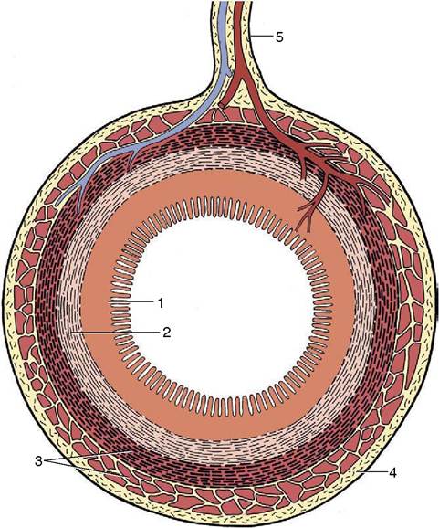
Figure 3-42 Transverse section through the gut. The artery and vein reach the gut via the mesentery; the larger branches fail to reach the antimesenteric border. 1, Mucosa; 2, submucosa; 3, muscle layer; 4, serosa; 5, mesentery.
when viewed in section. This explains the narrow streaks that are the common representation of the small intestine in radiographs obtained after the administration of a barium suspension.
Segmental and peristaltic movements continually alter the configuration in life.The intestine is composed of the usual four tunics (Figure 3-42). The luminal surface has a velvety appearance because of the innumerable tiny but densely packed projections known as the intestinal villi. These are fingerlike in the dog and horse but broader and leaflike in many species (Figure 3-43). In addition to the interspecific differences, variations in form and dimension may be present at different locations along the length of the small intestine. The appearance and the detailed morphology may be profoundly influenced by changes in diet (early weaning) or disease (microbial infections). The villi greatly increase the area of epithelium available for absorption; the efficiency of the process is enhanced by very generous subepithelial capillary plexuses (Figure 3-43, B). Microscopic intestinal glands (crypts) open to the surface between the bases of the villi. The crypts produce a mucous secretion, which coats the surface of the bowel, and various enzymes that contribute to the
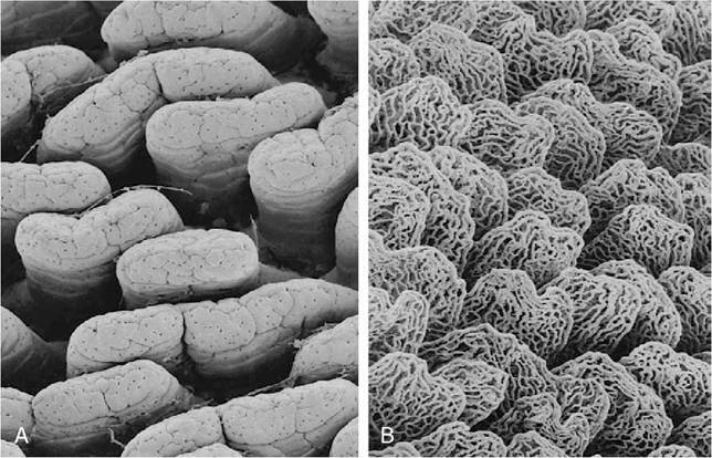
Figure 3—43 Scanning electron micrographs of rat duodenal villi (A) and of a vascular cast of the same tissue demonstrating Subepithelial capillary plexuses (B).
further digestion of carbohydrate and protein breakdown products.
Larger (Brunner’s) glands confined to the submucosa of the duodenum, especially its initial part, also secrete a protective mucus. A proportion of the cells lining the crypts, perhaps 1% of the total population, belong to the enteroendocrine (enterochromaffin) system (p. 222). Of several varieties, these cells form a series, commencing with the gastrin-producing cells of the stomach and extending through the small into the large intestine, that produces a number of hormones that influence various aspects of gastrointestinal activity. The intestinal components of the series, unlike that of the stomach, are under regulation by intrinsic nerves of the organ wall and largely outweigh the influence of the extrinsic nerve supply to the gut.
Cholecystokinin, which provokes contraction of the gallbladder, is an important member of the set.The great length and the villous surface of the small intestine combine to increase the absorptive area. In some species the absorptive area is also increased by permanent longitudinal and spiral folds; these are not pronounced in the dog, and the mucosal relief sometimes visible in radiographs is produced by temporary ridges.
The mucosa is rich in nodules of lymphoid tissue, both solitary and clumped; the larger aggregations (Peyer’s patches; [Figure 3-44]) cause visible depres-
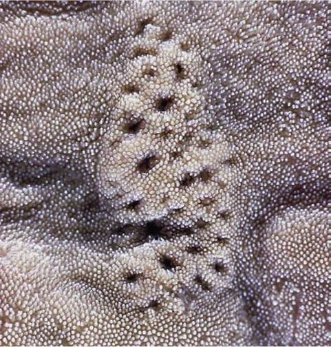
Figure 3-44 Patch of aggregated lymph nodules in ileum (horse).
sions and elevations of the mucosa that may become more obvious by the absence of a covering pile of villi. These aggregations tend to be more numerous and individually larger toward the junction with the large intestine.
Attention must be directed, however briefly, to the remarkable cycle of epithelial renewal exhibited by the lining of the small intestine throughout life. The epithelium is renewed by the mitotic division of cells in the depths of the crypts. The cells lining the crypts, continuously recruited in this way, gradually ascend to the surface, spread to embrace the bases of the villi, and continue up these to the summits where they are finally shed into the gut lumen. The passage from the bottom of a crypt to the summit of a villus takes about 3 days and involves a prodigious wastage—one calculation suggests a loss of about 1 g of epithelial cells for every centimeter stretch of the human small intestine every day. The process has the fortunate consequence of permitting rapid renewal of the integrity of the gut lining after extensive damage, such as the necrosis and loss by sloughing of the surface layer that occurs in certain infections in various domestic species. While repair is in train, the villi are reduced in size; they are not fully restored until a sufficiency of epithelial cells has again become available to clothe villi of normal height and proportions.
Both the liver and the pancreas discharge into the duodenum. The arrangement in the dog is for the bile duct and one pancreatic duct to discharge by separate openings on a (major duodenal) papilla a few centimeters beyond the pylorus, while the second larger pancreatic duct discharges on a smaller papilla a little farther on. Neither papilla is conspicuous.