THE SOUND-PERCEIVING APPARATUS
The inner ear has two main functions: the vestibular portion, which helps with position and balance, and the cochlear portion, which is responsible for hearing. The cochlear portion is connected to the cochlear nerve, a branch of the vestibulocochlear nerve (cranial nerve VIII), while the vestibular portion is connected to the vestibular nerve, another branch of the same cranial nerve.
The inner ear is housed in a bony structure called the osseous labyrinth, which is like a complex network of passages. The cochlea, responsible for hearing, is coiled to fit within the limited space available. If it were uncoiled, it would extend into the brain.10.4.1 Vestibular Structure and Function
The vestibular system helps us with balance and orientation. It’s located in the osseous labyrinth, specifically in the vestibule and three semicircular canals (front, side, and back). Each canal is positioned at right angles to the others. There’s also a cochlear part in the osseous labyrinth. Inside this labyrinth, there’s a membranous structure with fluid called endolymph, which is high in potassium and low in sodium. Another fluid, perilymph, is outside the membranous labyrinth. It can circulate with cerebrospinal fluid through the vestibular aqueduct, connecting to the brain’s subarachnoid space. This connection raises the risk of inner ear infections leading to meningitis (Figure 10.2).
In the vestibular portion, the membranous labyrinth has three semicircular ducts and two sacs called the utricle and saccule. The utricle connects to the saccule, and both connect to the cochlear portion. The cochlear sac, situated between layers of meninges, absorbs endolymph and allows immune cells to clear debris. Each semicircular canal has an enlarged section called an ampulla, housing equilibrium receptors called cristae. The utricle and saccule have sensory receptors called maculae, consisting of hair cells embedded in an otolithic membrane.
This membrane has a gelatinous material with otoliths, calcium carbonate crystals. Due to gravity, otoliths can stress hair cells, helping sense head position. The weight of otoliths also allows sensing linear head acceleration or deceleration. Rotational acceleration or deceleration is detected by the ampullary cristae in semicircular ducts. Hair cells in these structures respond to head movements and play a role in maintaining balance and orientation.10.4.2 Cochlear Structure and Function
The cochlea has a part called the cochlear duct or scala media, which runs through the center of the cochlea, dividing it into two parts: the scala vestibuli above and the scala tympani below. These parts are mostly separate, except for a small opening called the helicotrema at the tip of the cochlea. Inside the scala media, there are many structures known as organs of Corti. These structures convert sound waves into nerve impulses, which are then sent to the brain for us to hear. The position of each organ of Corti in the scala media, from the base (near the middle ear) to the top of the cochlea, and its individual nerve connection, determines the frequency of the sound we perceive (Figure 10.3).
Different frequencies of sound have different paths along the basilar membrane, which is the thicker base where nerve entries are located. A weak sound wave strengthens when it reaches the part of the basilar membrane with a resonant frequency similar to its own. High-frequency sounds travel a short
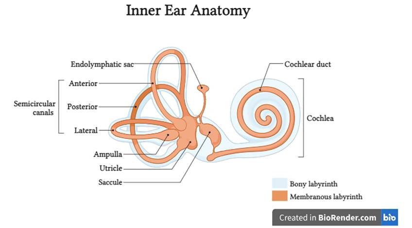
FIGURE 10.2 Structural component of the inner ear.
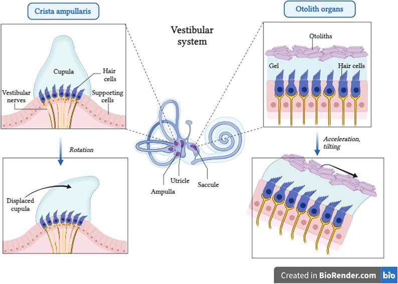
FIGURE 10.3 Structural component of the vestibular system of the inner ear.
distance before dying out, while low-frequency sounds travel a longer distance before doing the same. Every frequency between high and low is represented at different points along the basilar membrane. The organs of Corti are mainly made up of sensory receptors called hair cells with hairs reaching towards the tectorial membrane. When the basilar membrane vibrates due to sound waves, the hair cell cilia move against the tectorial membrane. This movement causes the hair cells to create a nerve impulse, which is then sent to the brain’s auditory cortex through the vestibulocochlear nerve (Figure 10.4).
10.4