THE SPINAL CORD
The spinal cord (medulla spinalis) is an elongated structure that is more or less cylindrical but with some dor- soventral flattening and certain regional variations in form and dimensions.
The most important of these are the thickenings (intumescentiae; Figure 8-15) of the parts that give origin to the nerves supplying the forelimbs and hindlimbs and the final caudal tapering (conus medullaris). The cord is divided into segments corresponding to the somites by the serial origins of the roots of the paired spinal nerves; the formation of these nerves has been described (p. 29). The relation of the segments to the vertebrae are considered in later chapters (see Figure 8-15).A simple transverse section shows a central mass of gray substance perforated in the midline by a small central canal, which is the residue of the lumen of the
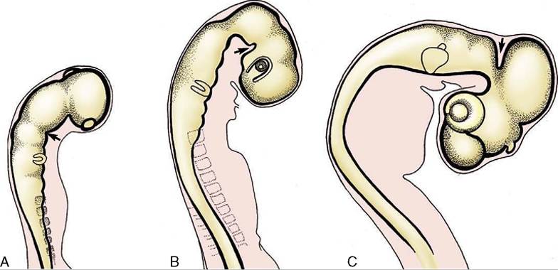
Figure 8-14 Formation of the caudal ventral (A), rostral ventral (B), and dorsal (C) flexures (arrows).
embryonic neural tube (see Figure 8-13). The gray substance, which has a crude resemblance to a butterfly or an H, is commonly described as exhibiting dorsal and ventral horns or columns; the former is a rather misleading term as the horns extend the length of the cord (Figure 8-16). The grayness is of course produced by the restriction of the perikarya to this part. The dorsal horn corresponds to the alar plate. It contains somatic afferent neurons dorsomedially and visceral afferent neurons dorsolaterally (see Figure 8-17). The ventral horn corresponds to the basal plate; it is composed of somatic efferent neurons, which are located ventrally, and visceral efferent neurons, which form an additional lateral horn confined to the thoracolumbar and sacral regions of the cord.
The neurons within each horn are more specifically grouped according to their functional and topical associations, but this is not grossly discernible.
The white substance that envelops the gray is divided into three funiculi on each side (Figure 8-18H,II,III). The dorsal funiculus is contained between a shallow dorsal sulcus, extended deeply by a median glial septum,
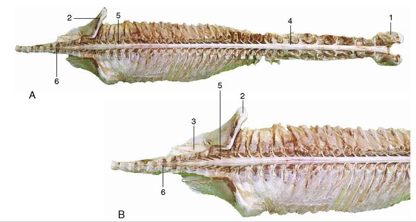
Figure 8-15 A, Dorsal view of the spinal cord and the vertebral pedicles of the horse. The spinal cord is shorter than the vertebral canal (ascensus medullae spinalis). B, Enlargement of the caudal part. 1, Atlas; 2, ilium; 3, sacrum; 4, cervical intumescence; 5, lumbar intumescence; 6, cauda equina.
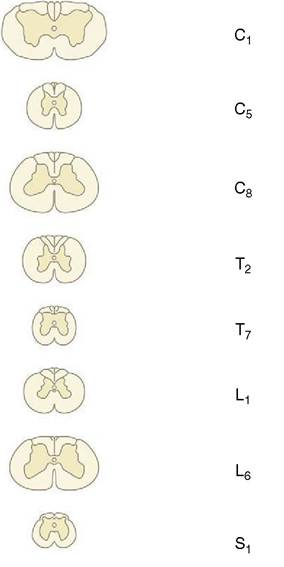
Figure 8-16 Transverse sections of the canine spinal cord (the levels are indicated). Note the changes in diameter of the cord and in the relative proportions of gray and white substance.
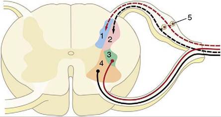
Figure 8-17 Schematized subdivision of the gray substances in the spinal cord. 1, Somatic afferent neurons; 2, visceral afferent neurons (1 and 2 form the dorsal horn); 3, visceral efferent neurons; 4, somatic efferent neurons (3 and 4 form the ventral horn); 5, dorsal root ganglion.
and the line of origin of the dorsal roots of the spinal nerves (see Figure 8-13). The lateral funiculus is contained between the lines of the dorsal and ventral roots, and the ventral funiculus is contained between the line of the ventral roots and a ventral fissure that penetrates far into the white substance, although it leaves a considerable commissure connecting the right and left halves. This ventral fissure is occupied by a mass of pia that appears as a glistening streak on the surface of the cord.
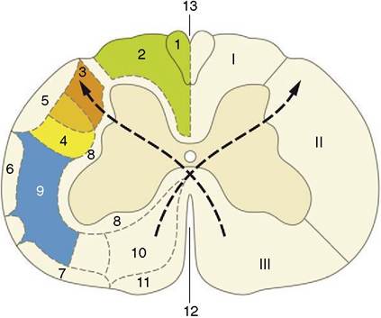
Figure 8-18 Hypothetical transverse section of the canine spinal cord showing the location of some principal tracts.
The curved arrows indicate the crossing of the pyramidal tracts. (The drawing has been simplified for the sake of clarity.) I, Dorsal funiculus; II, lateral funiculus; III, ventral funiculus. 1, Fasciculus gracilis; 2, fasciculus cuneatus; 3, lateral corticospinal tract; 4, rubrospinal tract; 5, dorsal spinocerebellar tract; 6, ventral spinocerebellar tract; 7, spinoolivary and olivospinal tracts; 8, propriospinal system (fasciculi proprii); 9, spinothalamic tract; 10, ventral corticospinal tract; 11, vestibulospinal tract; 12, ventral median fissure; 13, dorsal median sulcus.The funiculi are composed of ascending and descending nerve fibers, of which many are grouped within bundles (fasciculi or tracts) of common origin, destination, and function (see Figure 8-18). Certain of these are mentioned later.