THE SPLEEN
The form of the spleen (see also p. 264) is the same in dogs and cats; it is an elongated, roughly dumbbellshaped organ that lies more or less vertically against the left abdominal wall (Figure 14-7, A/4).
Its position is much influenced by the distention of the stomach (and by its own capacity to become engorged). The dorsal end reaches the left crus of the diaphragm, passing between the gastric fundus and the cranial pole of the left kidney under cover of (usually) the last two ribs.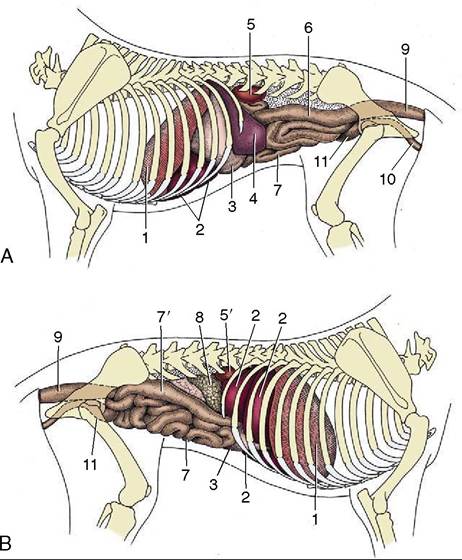
Figure 14-7 Visceral projections on the left (A) and right (B) canine abdominal walls. 1, Diaphragm; 2, liver; 3, stomach; 4, spleen; 5, 5', left and right kidneys; 6, descending colon; 7, small intestine; 7, descending duodenum; 8, pancreas; 9, rectum; 10, female urogenital tract; 11, bladder.
The larger ventral end may cross the ventral midline, to reach under the costal cartilages of the right side. It then provides a dense triangular shadow on the abdominal floor in lateral radiographs (Figure 14-8, AJ3∖ A similar shadow between the stomach and left kidney may reveal the position of the organ in ventrodorsal films. In the cat, the ventral part of the spleen is always located outside the rib cage. The parietal surface makes contact (in dorsoventral sequence) with the diaphragm, costal arch, and abdominal muscles. The visceral surface is divided by a hilar ridge into a cranial strip related to the stomach and a caudal strip related to the left kidney and intestine.
The wide gastrosplenic ligament attaches the spleen to the greater curvature of the stomach. Although permitted considerable mobility, the spleen follows the movements of the stomach. When the stomach enlarges, the spleen is displaced caudally and ventrally, reaching the pelvic inlet; it may then be palpated through the abdominal wall.
Another restraining influence is provided by the tether of its blood vessels. The splenic artery and vein pass (as several divergent branches) to the dorsal end of the spleen. The splenic artery arises as a branch of the celiac artery, and before reaching the spleen, it gives off branches to the left limb of the pancreas. The left gastroepiploic vessels are detached about the middle of the hilus and cross to the greater curvature of the stomach within the gastrosplenic ligament (Figure 14-9/3,77). The splenic lymph nodes lie by the splenic vessels, a few centimeters distant from the organ. The spleen has efferent (which follow large arteries) but no afferent lymphatic vessels.
The spleen serves as an important blood reservoir in the dog and cat, and its size and weight therefore vary widely (Figure 14-6). The spleen in a resting dog or cat contracts and relaxes rhythmically because of the presence of many smooth muscle fibers throughout the organ. These fibers relax when anesthetics are used, which results in marked splenic enlargement, and contract because of stress or injection of catecholamines, expelling free blood cells and plasma from the red pulp. The spleen has no parasympathetic nerve supply.
Rupture of the spleen is not uncommon after traffic accidents, but fortunately the organ may be removed without risk to life. The relatively loose attachment of the spleen to the stomach facilitates access to the vascular supply at surgery (splenectomy).
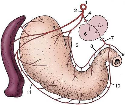
Figure 14-9 The blood supply of the stomach and spleen, caudal view; schematic. 1, Aorta; 2, celiac a.; 3, splenic a.; 4, hepatic a.; 5, left gastric a.; 6, indication of the liver; 7, gastroduodenal a.; 8, right gastric a.; 9, cranial pancreaticoduodenal a.; 10, right gastroepiploic a.; 11, left gastroepiploic a.
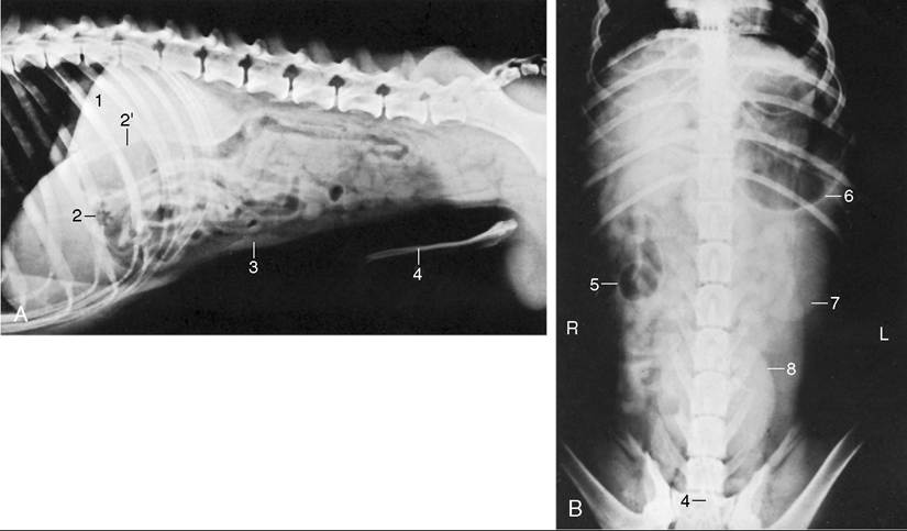
Figure 14-8 Lateral (A) and ventrodorsal (B) radiographic views of the canine abdomen. 1, Liver; 2, pyloric part of stomach; 2', descending duodenum; 3, spleen; 4, os penis; 5, cecum; 6, fundus of stomach; 7, left kidney; 8, bladder.