THE SPLEEN
The spleen lies within the left dorsal part of the abdomen where it is largely, if not wholly, protected by the most
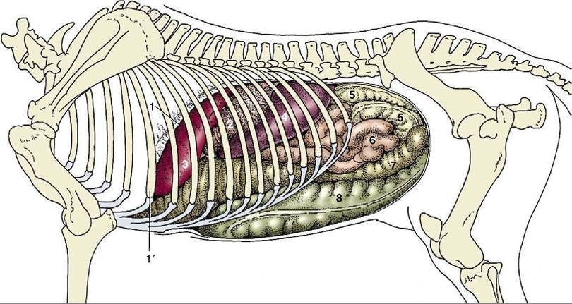
Figure 21-6 Visceral projections on the left abdominal wall (including the diaphragm).
1, Cut edge of diaphragm; Γ, rib 6; 2, stomach; 3, liver; 4, spleen; 5, descending colon (banded); 6, jejunum (smooth); 7, left dorsal colon; 8, left ventral colon.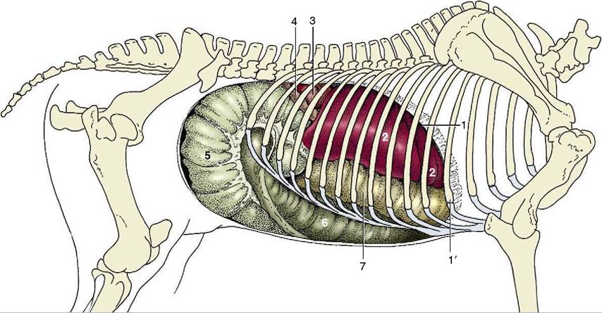
Figure 21-7 Visceral projections on the right abdominal wall (including the diaphragm). 1, Cut edge of diaphragm; Γ, rib 6;
2, liver; 3, right kidney; 4, descending duodenum; 5, body of cecum; 6, right ventral colon; 7, right dorsal colon.
caudal ribs, from which it is separated only by the diaphragm. The broad dorsal base lies under the last three ribs, although a small corner may project against the flank. The pointed ventral apex reaches forward to about the ninth or tenth rib, a handbreadth above the costal arch (Figure 21-6/4). The cranial margin is concave, the caudal margin is convex, and the organ is thus approximately sickle-shaped. The parietal surface is generally smooth, though sometimes marked by depressions that may even perforate to the visceral surface. It lies against the diaphragm but is not joined to this. The visceral surface presents three parts. A small dorsal region fits against the left crus of the diaphragm and left kidney and is bound to these by phrenicosplenic and renosplenic ligaments (Figure 21-8/6,7). The remainder of the visceral surface is divided by a ridge along which the splenic artery runs and to which the greater omentum attaches. The narrow strip cranial to the ridge, the gastric surface, is applied to the greater curvature of the stomach (see Figure 21-21); the larger
2
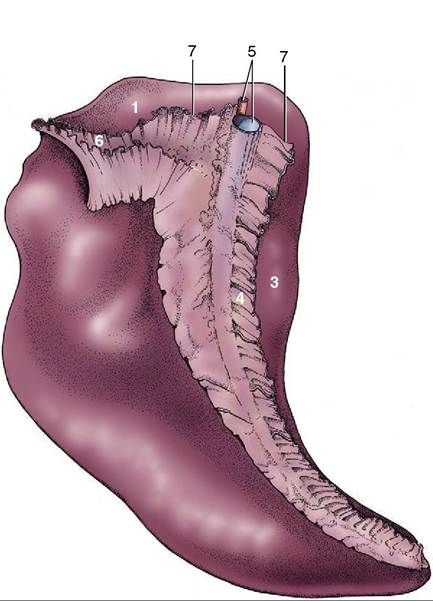
Figure 21-8 Visceral surface of the spleen.
1, Renal surface; 2, intestinal surface; 3, gastric surface; 4, greater omentum (gastrosplenic ligament); 5, splenic artery and vein; 6, reno- splenic ligament; 7, phrenicosplenic ligament.area caudal to the ridge, the intestinal surface (Figure 21—21/1), is related to various parts of the intestinal mass.
The thick capsule contains a considerable amount of smooth muscle, which allows much variation in volume because the spleen becomes engorged when the capsule is relaxed. This occurs in certain diseases and is very obvious in animals that have succumbed to anthrax. The organ is steel blue on first removal from the fresh carcass but turns reddish brown on exposure to the air. This color is derived from the red pulp that forms the bulk of the parenchyma. The white pulp that flecks the red is not normally visible to the naked eye.
The position of the spleen naturally varies with respiration. Usually only the caudal margin is within reach on rectal exploration (see Figure 22—23/10); a greater part becomes accessible when the stomach is distended.