The Starling Equation Quantifies the Interaction of Oncotic and Hydrostatic Forces Acting on Water
The following equation expresses mathematically the interaction between osmotic pressures and hydrostatic pressures in determination of the net force (net pressure) acting on water.
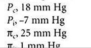
Nominal values for each pressure are also listed:
where Pi is capillary hydrostatic pressure, P1 is interstitial fluid hydrostatic pressure, πc is capillary plasma oncotic pressure, and πi is interstitial fluid oncotic pressure. Nominal values for systemic capillaries are as follows:
,∙1> ∙................. *©
The solution of this equation, with nominal values inserted for each term, follows:
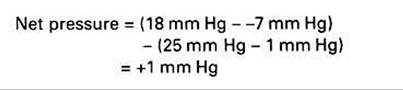
A positive net pressure favors filtration (a negative net pressure would indicate that reabsorption is favored). The small magnitude of the net pressure (1 mm Hg) indicates that the hydrostatic and osmotic forces that affect water are almost in balance (i.e., there is only a slight tendency for filtration). The quantitative analysis of how oncotic and hydrostatic pressures affect water movement across capillary walls was first derived by Ernest Henry Starling (the same scientist for whom Starling’s law of the heart is named). Therefore the oncotic and hydrostatic pressures that act on water are often called Starling forces. Furthermore, the tendency for the net oncotic effect to be closely balanced by the net hydrostatic effect is often referred to as the balance of Starling forces. Starling realized that the actual rate of water movement across capillary walls is affected both by the magnitude of the imbalance between hydrostatic and oncotic forces and by the permeability of the capillary wall to water.
These ideas are expressed in the following equation, which indicates that the movement of water is equal to the permeability of the capillary wall (given as the filtration coefficient Kf) multiplied by the net difference between the hydrostatic and oncotic pressures:
Examination of this equation reveals that the tendency for the filtration of water out of capillaries can be enhanced by (1) increasing the hydrostatic difference between capillary blood and interstitial fluid, (2) decreasing the osmotic tendency for water to be reabsorbed, or (3) increasing the permeability of the capillary to water (i.e., increasing the filtration coefficient).
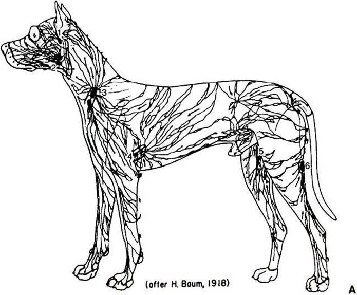
FIGURE 23-4 Anatomical (A) and schematic (B) overviews of the lymphatic system. The lymphatic vessels collect excess interstitial fluid from tissues throughout the body (including the lungs) and carry it to the subclavian veins, where the lymph reenters the bloodstream. Lymph moves through lymph vessels via bulk flow.The driving force for this flow is interstitial fluid hydrostatic pressure minus subclavian vein pressure. Lymph flow is also promoted by the massaging action exerted on lymph vessels by contraction and relaxation of skeletal muscles and (in the lungs) by the pressure variations accompanying inspiration and expiration.The lymph vessels contain one-way valves, which prevent the backflow of lymph.Thus, massaging actions propel lymph in one direction only: toward the subclavian vein. In addition, some lymph vessels have smooth muscle in their walls, and the alternating contraction and relaxation of this smooth muscle also propels lymph flow toward the subclavian veins.The numbers in A identify the major lymph nodes.The magnified inset n B depicts the typical, net filtration of fluid out of a blood capillary and into the interstitial space.This excess interstitial fluid is collected and carried away by the lymph capillaries.Three red blood cells are depicted in the blood capillary. Plasma is indicated in yellow, interstitial fluid and lymph in blue. (A from Getty R: Sisson and Grossman's the anatomy of the domestic animal, vol 2, Philadelphia, 1975, Saunders.)