THE STOMACH
The stomach, interposed between the esophagus and small intestine, is the dilated part of the digestive tract in which the processes of digestion are initiated. It is succeeded by the intestine, which consists of a proximal small intestine (the principal organ of digestion and absorption in most species) and a distal large intestine (generally much shorter and especially concerned with the dehydration of the food residue).
However, among mammals there exists considerable diversity in the form and structure of these two parts of the digestive system, which are closely associated in function and which are collectively known as the gastrointestinal tract. Much of this diversity is clearly adaptive and reflects the habitual diet of the various groups. The concentrated diet of carnivores is most easily digested, and these animals have a small and simple stomach (Figure 3-35, A) and a relatively short and uncomplicated intestine. The fodder of herbivores is less easily managed; it has a lower nutritive value and must be consumed in large amounts. Moreover, a major part consists of celluloses and other complex carbohydrates that are not susceptible to the action of mammalian digestive enzymes. These substances can be utilized only if they are first broken down by symbiotic microorganisms; this is a relatively slow process that requires the provision of a large fermentation chamber where food may be held in an environment favorable to the multiplication and activity of the microorganisms. In some herbivorous species, such a chamber is supplied by a greatly enlarged and subdivided stomach, in others
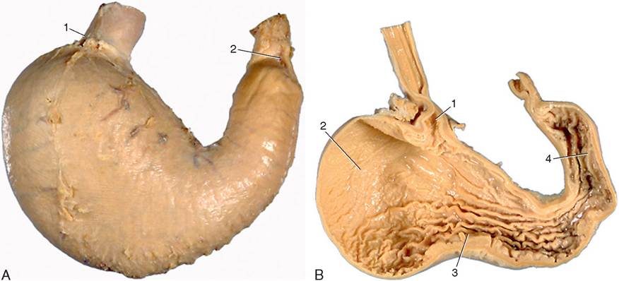
Figure 3-35 A, Visceral surface of stomach (dog). 1, cardia; 2, pylorus. B, Interior of stomach (dog). 1, cardiac opening; 2, fundus; 3, body; 4, pyloric antrum.
by a voluminous and complicated large intestine. Ruminants illustrate the first alternative, the horse the second. Some indication of the range of variation of gastrointestinal anatomy among domestic species is provided by Figure 3-36. Detailed accounts are found in the chapters concerned with individual species; the description that follows is largely confined to the simple organs of the dog and cat.
The stomach (ventriculus) receives food from the esophagus and retains it for a time before discharging it into the duodenum, the first part of the small intestine. The stomach of the dog has a relatively modest capacity, ranging from 0.5 to 6.0 L according to breed, and conforms to a pattern that is common to most carnivores and indeed to many other mammals, including ourselves. It consists of two distinct parts that converge and join at a ventral angle (Figure 3-37). The larger part, into which the esophagus opens at the cardia, lies mainly to the left of the median plane, well forward under cover of the ribs and in direct contact with the liver and the diaphragm; it is relatively distensible and rapidly expands to accommodate a meal. The second part is narrower, has thicker walls, and is more constant in appearance since it is less affected by the presence of a meal; it passes to the right to continue into the duodenum at the pylorus (Figure 3-35, B). The cranial (parietal) aspect of both parts is mainly in contact with the liver, while the more numerous relations of the caudal (visceral) surface include the intestinal mass, left kidney, pancreas, and greater omentum. The left part of the margin is applied to the hilar region of the spleen.
Other terms are available when it is necessary to refer to particular regions of the stomach more precisely. The large left sac is divided between a blind dome (fundus) rising above the cardia and a body (corpus) extending from the cardia to the ventral angle. The more tubular right or pyloric part is divided between a more proximal pyloric antrum and a more distal pyloric canal; the distinction is based on the terminal muscular thickening (see Figure 3-35, B).
The margin separating the two surfaces is divided between greater and lesser curvatures, each of which runs between the cardiac and pyloric openings. The convex greater curvature gives attachment to the greater omentum, of which a part (gastrosplenic ligament) connects the spleen with the stomach. The shorter, concave lesser curvature is connected with the liver by the lesser omentum. This curvature is marked by a sharp change in direction known as the angular notch (incisura).The stomach wall is composed of layers corresponding to those of the esophagus and intestine. The external peritoneum or serosa covers the entire organ, adhering to the underlying muscle, except along the curvatures, where it is reflected to continue into the omenta; its absence from the curvatures makes them the parts most likely to burst when the organ is excessively distended.
The next coat is of smooth muscle and is arranged in three layers, each of which is incomplete but with its deficiencies compensated by the others. The external layer is more or less longitudinal and continues the outer muscle of the esophagus; it is concentrated along the curvatures, although it spreads more widely over the
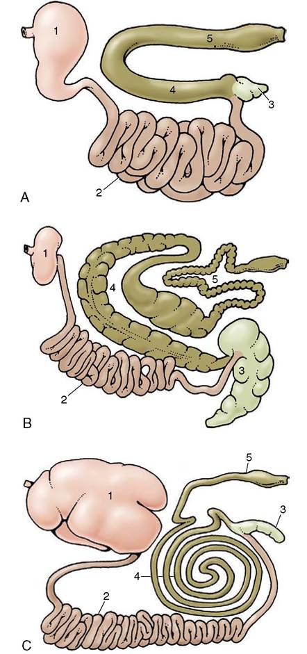
Figure 3-36 Gastrointestinal tracts of the dog (A), of the horse (B), and of cattle (C) laid out in one plane. 1, Stomach; 2, small intestine; 3, cecum; 4, ascending colon; 5, descending colon.
pyloric part. The middle layer is disposed in hoops, and those most proximal form a weak sphincter around the cardia; beyond this the pattern is interrupted by the projection of the fundus, but it is resumed at a lower level. It then continues to the pyloric canal, where the hoops are bunched together on the lesser curvature, forming a muscular knot (that in some species produces an obvious projection into the lumen) and fanning out on the greater curvature; the edges of this “fan” are sometimes held to constitute proximal and distal pyloric sphincters.
The innermost layer is very incomplete but compensates for the deficiencies in the circular muscle; particularly stout fascicles arch above the cardia before continuing distally to each side of the lesser curvature, extending toward, but not beyond, the angular notch (see Figure 3-37).The thin submucosa internal to the muscle is separated from the mucosa proper by a plexiform muscularis mucosae. It contains major arterial and venous plexuses and also a wealth of elastic fibers that help the muscularis mucosae throw the mucosa of the empty organ into the folds (rugae) that provide the characteristic surface relief (Figure 3-37 and Figure 3-38, A). These folds are predominantly longitudinal in orientation, although individually tortuous; they are completely effaced only when the stomach is grossly distended.
The entire gastric mucosa is densely pockmarked by innumerable tiny depressions. These so-called gastric pits (many would be better described as crevices) are invisible to the naked eye but account for the surface folding seen in histological sections (Figure 3-38, B). The surface epithelium of columnar, mucus-secreting cells continues into the pits and even extends into the uppermost parts of the gastric glands that deliver their products into the depth of the pits. This epithelium is largely responsible for the protective coat that makes gastric mucosa slimy to the touch. The gastric glands are of three varieties, termed cardiac, proper gastric (fundic), and pyloric, although it must be stressed that in many species, including the dog, their distribution does not exactly coincide with the gross regions that bear the same names. The cardiac and pyloric glands produce additional mucus, whereas the proper gastric glands are alone responsible for the gastric juice active in digestion by virtue of its pepsin and hydrochloric acid content. The enzyme is the product of its most numerous (chief) cell type, the acid of the fewer parietal cells; there is also a further contingent of mucus-secreting cells.
It is claimed that the proper gastric glandular region has a somewhat darker hue than the remainder of the mucosa.The blood supply to the stomach comes from all three chief branches of the celiac artery and is particularly generous along the two curvatures (Figure 3-39). The arteries anastomose quite freely externally and also within the stomach wall. For the most part, the arteries that penetrate the wall pass to the submucosa before branching to form an elaborate plexus from which both the muscular and the mucosal coats are fed. The mucosal branches supply unusually wide-bored capillaries below the epithelium and about the glands.
The veins are similarly arranged and ultimately combine to form trunks that join the portal vein. Numerous arteriovenous anastomoses provide a means
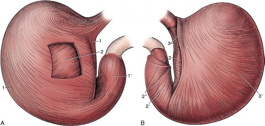
Figure 3-37 The tunica muscularis of the canine stomach. A, Parietal surface after removal of the serosa. B, Stomach turned inside out with the mucosa removed. The tunica muscularis comprises outer longitudinal, middle circular, and inner oblique layers. The longitudinal layer clothes the curvatures (1) and the pyloric part (1') but is thin over the body. The circular layer surrounds the body (2) and is especially prominent on the pyloric part (2'), where it furnishes the pyloric sphincters (2"). The oblique layer (3) is thickest along the lesser curvature, where it forms two lips that fuse over the cardia (cardiac loop); it is thin where it lines the fundus and body (3').
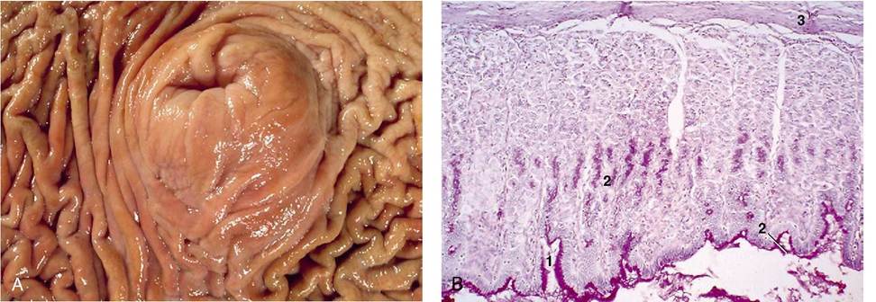
Figure 3-38 A, Protruding cardia surrounded by longitudinal folds. B, Mucosa of stomach (PAS-H; 70?) (dog). 1, gastric pit; 2, mucopolysaccharide-secreting cells; 3, lam. muscularis mucosae.
of regulating mucosal blood supply, and much blood is diverted from the capillary bed of the fasting organ.
Lymph vessels are present in profusion, particularly in the submucosa.
They lead to several gastric nodes, each charged with the drainage of a particular territory.The stomach is innervated by parasympathetic fibers within the two vagal trunks and by sympathetic fibers that reach the organ with the arteries. The efferent fibers of both sets are accompanied by more numerous afferent fibers. Parasympathetic fibers of the vagus synapse on ganglion cells in intramural plexuses within the submucosa and between the muscle coats and exert a high measure of control over gastric motility. The effects of vagal stimulation on the proximal and distal regions of the stomach are dissimilar: in the proximal stomach, vagal activity suppresses muscular contrac-
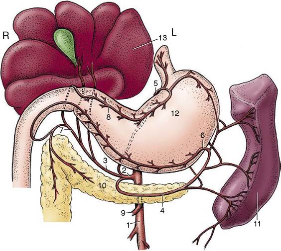
Figure 3-39 Distribution of the celiac artery of the dog (ventral view). 1, Aorta; 2, celiac artery; 3, hepatic artery; 4, splenic artery; 5, left gastric artery; 6, left gastroepiploic artery; 7, gastroduodenal artery; 8, right gastric artery; 9, cranial mesenteric artery; 10, pancreas; 11, spleen; 12, stomach; 13, liver.
tion and leads to adaptive relaxation, whereas in the distal stomach, vagal stimulation causes intense peristaltic activity. Vagal stimulation of distal antral motility is mediated by acetylcholine, but the identity of the inhibitory mediator is not well established; it may be vasoactive intestinal peptide. The intramural plexuses are involved in the local reflexes in which the stomach wall reacts to direct stimulation. Sympathetic and parasympathetic fibers also innervate the surface epithelium and glands, but only parasympathetic fibers end on the intragastric endocrine cells. Division of the vagal nerves, either the main trunks or selected branches, reduces gastric activity and secretion.
The topography and the form of the living stomach of the dog are much influenced by functional changes. The empty stomach is small and contracted toward the fixed point of the esophageal entrance. It lies entirely within the rib cage and fails to reach the abdominal floor. The wall is generally inert except for occasional weak peristaltic contractions, and little secretion from the glands occurs. Any residual peristaltic activity ceases as soon as food is offered (or anticipated). Secretion increases as a reflex response to the taste of food or the effort of mastication; it appears to be independent of food actually reaching the stomach. When food does arrive, it first collects in layers (because as yet no mixing movements are present) and largely occupies the body, which expands in all directions but principally ventrally and caudally. A motor response is delayed, and when it begins, it is relatively slow in building to a peak. Peristaltic contractions commence near the cardia and course distally, accelerating and becoming more vigorous when they reach the muscular pyloric antrum. The terminal segment contracts en masse, and the injection of ingesta into the duodenum therefore occurs when the wave is still some distance from the pylorus. Radiographic studies suggest that the pylorus is open for about one third of the time; it is probable that emptying is more dependent on intermittent increase of the intra- gastric pressure than on the regular peristaltic activity.
The effects of feeding on topography and relations are considerable, especially in animals kept under regimens that allow them to feed seldom but to repletion. The fully distended stomach may extend almost to the umbilicus—or even beyond this in the puppy—pushing the intestinal mass dorsally and caudally. The liver is pushed to the right while the spleen, tethered to the left
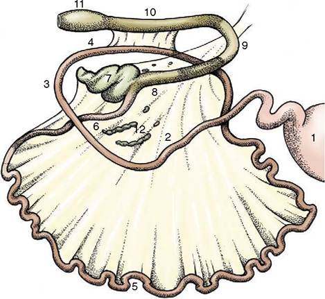
Figure 3-40 Intestinal tract of the dog (schematic). 1, Stomach; 2, descending duodenum; 3, caudal flexure; 4, ascending duodenum; 5, jejunum; 6, ileum; 7, cecum; 8, ascending colon; 9, transverse colon; 10, descending colon; 11, rectal ampulla; 12, jejunal lymph nodes.
part of the greater curvature, follows the expansion of that side of the stomach.