The Stomach
The stomach is of the simple type, presenting fundus, corpus, and a pyloric part (Fig. 34.8/2). The first two are generally confined to the left side of the abdomen but may extend across the median plane when the stomach is grossly distended.
They are cranially related to the liver and diaphragm. The pyloric part extends to the right and is also in contact with the liver. All parts are related caudally to various parts of the intestinal mass, with the principal relation being to the ascending colic spiral. It is only when grossly distended that the stomach makes contact with the abdominal floor and, on the left, extends beyond the protection of the rib cage. A feature unique to the pig among domestic species is the presence of a conical diverticulum (Fig. 34.8/2) projecting caudally from the fundus.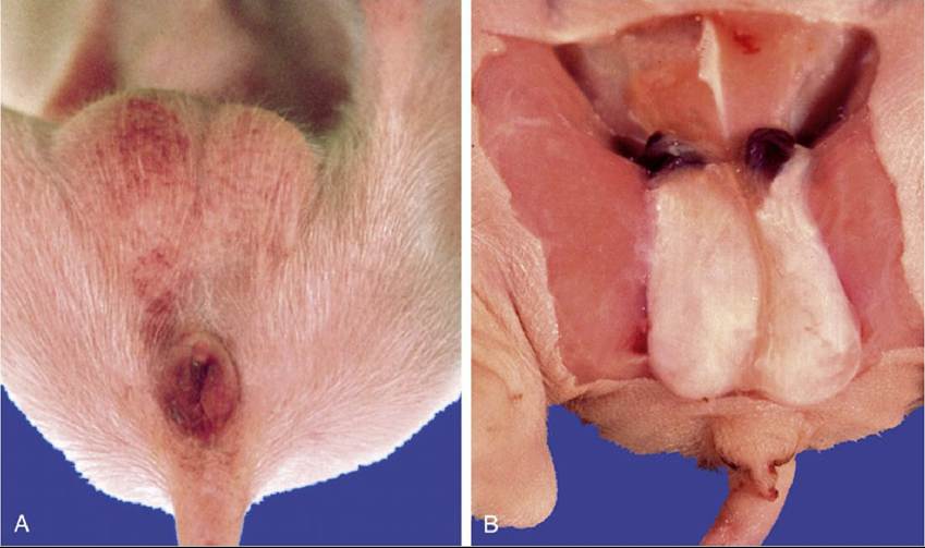
FIG. 34.3 A, Gubernacula in a freemartin piglet. B, Exposed.
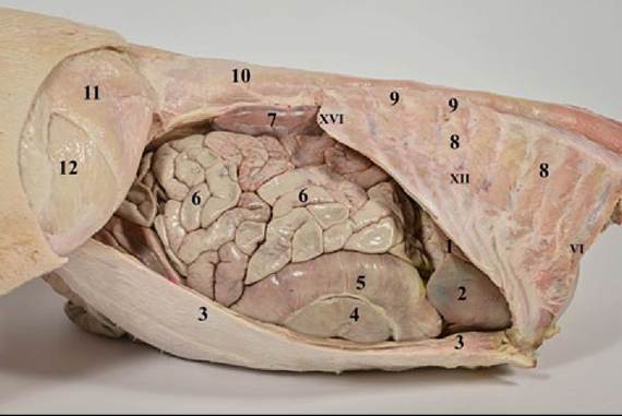
FIG. 34.4 Abdominal cavity of the pig from the right side. 1, Liver, right lateral lobe; 2, liver, right medial lobe; 3, abdominal muscles (cut); 4, ascending colon (gyri centrifugales); 5, ascending colon (gyri centripetales); 6, jejunal loops; 7, right kidney; 8, intercostal muscles; 9, serratus dorsalis muscle (caudal part); 10, epaxial muscles (iliocostalis lumborum and longissimus); 11, tensor fasciae latae muscle; 12, vastus lateralis muscle; VI, XII, XVI: ribs with respective numbers.
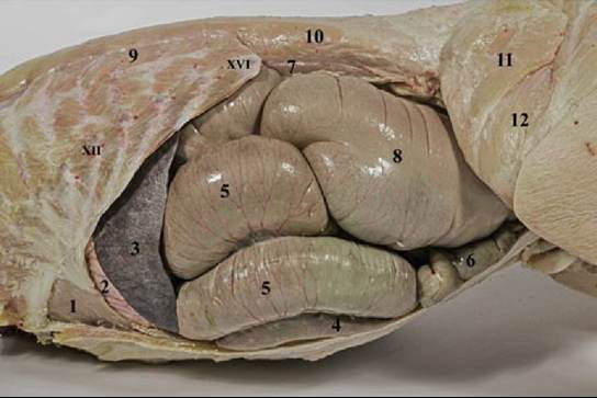
FIG. 34.5 Abdominal cavity of the pig from the left side. 1, Liver, left lateral lobe; 2, stomach; 3, spleen;
4, ascending colom (gyri centrifugales); 5, ascending colon (gyri centripetales); 6, jejunal loops; 7, left kidney; 8, cecum; 9, serratus dorsalis muscle (caudal part); 10, epaxial muscles (iliocostalis lumborum and longissimus); 11, tensor fasciae latae muscle; 12, vastus lateralis muscle XII, XvI: ribs with according numbers.
The interior displays a narrow nonglandular strip of mucosa that extends into the diverticulum and follows the lesser curvature for some distance below the cardia (Fig. 34.9/1). The remainder of the mucosa is divided into the usual three glandular regions, which are more clearly distinguished by color than in most species, although their borders are not always sharply defined (Fig. 34.9/2a, 2b, and 2c). A second feature of distinction is the very prominent torus narrowing the pyloric canal at the exit into the duodenum (Fig. 34.8/10).
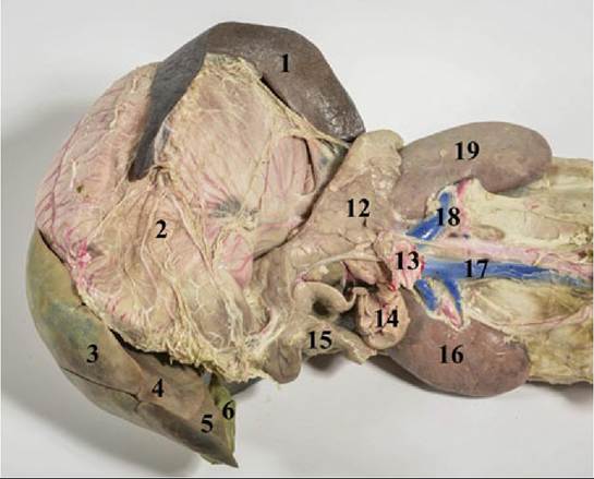
FIG. 34.6 Abdominal organs of the pig, after removal of the intestines. 1, Spleen; 2, stomach; 3, liver, left medial lobe; 4, liver, left lateral lobe; 5, liver, quadrate lobe; 6, gallblader; 12, pancreas, left lobe; 13, cranial mesenteric artery (cut); 14, pancreas, right lobe; 15, descending duodenum; 16, right kidney; 17, caudal vena cava; 18, renal artery and vein; 19, left kidney.
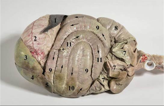
FIG. 34.7 Abdominal cavity of the pig (ventral view). 1, Spleen; 2, stomach; 3, liver, left lateral lobe; 4, liver, left medial lobe; 5, liver, quadrate lobe; 7, jejunal loops; 8, cecum; 9, ascending colon (gyri centripetales); 10, ascending colon (flexura centralis); 11, ascending colon (gyri centrifugales). Asterisk shows the teres hepatis ligament at the attachment to the adbominal wall at the navel.
Although the omenta are arranged much as in the dog, the greater one is less extravagantly developed, does not intervene between the intestines and the abdominal floor, and is therefore not encountered when the abdomen is first opened.
The arrangement of abdominal viscera is demonstrated in transverse sections taken at the 11th (Fig. 34.10) and 16th (Fig. 34.11) thoracic vertebrae.
FIG. 34.9
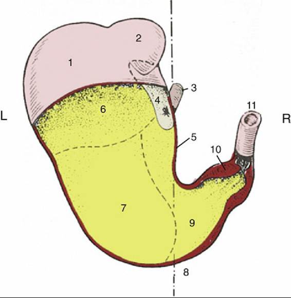
FIG. 34.8 Stomach partially opened, Caudoventral view, semischematic. 1, Fundus; 2, diverticulum; 3, esophagus; 4, nonglandular mucosa; 5, lesser curvature; 6, cardiac gland region; 7, region of proper gastric glands; 8, approximate position of median plane; 9, pyloric gland region; 10, torus pyloricus; 11, duodenum.
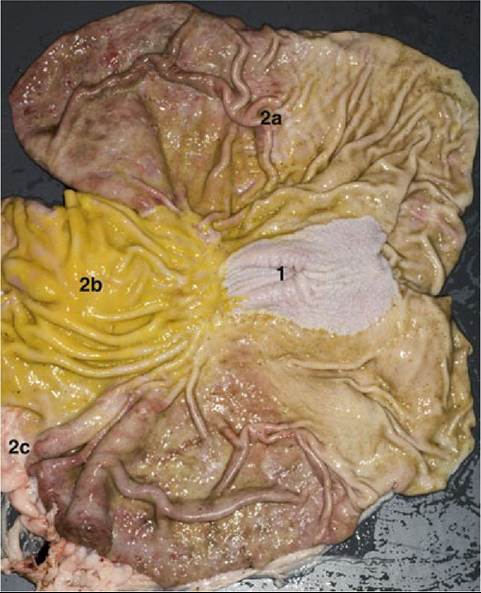
The stomach laid open (cardia to the right). 1, Nonglandular region; 2a, region with cardiac
glands; 2b, region with proper gastric glands; 2c, region with pyloric glands.