The Structure of the Glomerulus Allows Efficient, Selective Filtration
The glomerular tuft is composed of a network of capillaries (Figure 41-2). In mammals, blood from the renal artery is delivered to the afferent arteriole, which divides into numerous glomerular capillaries.
The capillaries coalesce to form the efferent arteriole, which conducts the filtered blood away from the glomerulus (Figure 41-3). Birds have three pairs of renal arteries: anterior, middle, and posterior. Avian kidneys con-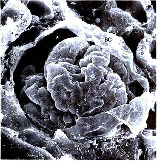
FIGURE 41-2 Scanning electron micrograph of rat glomerulus.The glomerular tuft is a complex network of capillaries that is encased in visceral epithelial cells and Bowman's capsule (BC). Between the visceral epithelial cells and BC is Bowman's space (asterisk), where the glomerular filtrate is collected and delivered to the proximal tubule. (Magnification "600.)
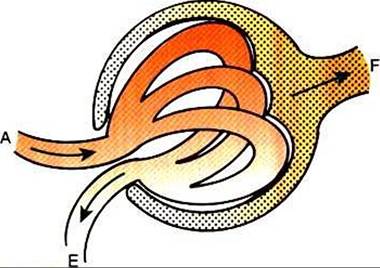
FIGURE 41-3 Schematic illustration of glomerulus.The afferent arteriole (A) carries blood to the glomerulus and subdivides into numerous glomerular capillaries. Water and solutes cross the glomerular capillary wall into Bowman's space, forming the glomerular filtrate (F, stippled area), which flows into the proximal tubule.The glomerular capillaries coalesce, and the filtered blood leaves the glomerulus through the efferent arteriole (Eλ
tain both mammalian-type and reptilian-type nephrons; in glomeruli of reptilian-type nephrons, the capillaries have few branches.
The glomerular tuft is encased within Bowmans capsule, which is lined with a single layer of epithelium. The area between the glomerular tuft and Bowmans capsule is known as Bowman's space and is the site of collection of the glomerular filtrate, which is tunneled directly into the first segment of the proximal tubule.
'Γhe structure of the glomerular capillaries is important in determining the rate and selectivity of glomerular filtration. The wall of the capillary consists of three layers: the capillary endothelium, the basement membrane, and the visceral epithelium (Figure 41-4). The capillary endothelium is a single layer of cells with cytoplasmic extensions that are pierced by fenestrae (windows). The endothelial fenestrae provide channels for the passage of water and Iioncellular components from the blood to the second layer of the glomerular capillary wall, the glomerular basement membrane. This is an acellular structure composed of various glycoproteins, including type IV and type V collagens, proteoglycans, laminin, fibronectin, and entactin. The glomerular basement membrane is arranged in three layers, presumably created during development by the fusion of the basement membranes of the endothelial and epithelial cell layers. The three layers are named according to both their density with respect to an electron beam and their relative position. As shown in Figure 41-4, the lamina densa (dense layer) is relatively dark because it is relatively resistant to the passage of electrons when viewed with a transmission electron microscope. The lamina densa is composed of tightly packed glycoprotein fibrils. It is sandwiched between the lamina rara interna (inside thin layer) on the endothelial side of the glomerular basement membrane and the lamina rara externa (outside thin layer) on the epithelial side of the glomerular basement membrane. The laminae rarae arc composed of a loose network of glycoprotein fibrils.
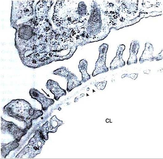
FIGURE 41-4 Transmission electron micrograph of rat glomerular capillary wall.The three main layers of the capillary wall are viewed in cross section. A single layer of glomerular capillary endothelial cells lines the capillary lumen (CL). Numerous fenestrae (arrowheads) pierce the endothelial cells.
On the outside of the capillary is a single layer of visceral epithelial cells. At the top of the micrograph is a portion of the cell body of a visceral epithelial cell. The secondary foot processes are aligned along the capillary wall, and the spaces between them are spanned by the slit diaphragm (arrow). Between the endothelial and epithelial cell layers is the glomerular basement membrane, consisting of the electron-lucent lamina rara interna adjacent to the endothelial cells, the lamina densa (asterisk), and the lamina rara externa adjacent to the visceral epithelial cells. (Magnification "19,000.)The third compartment of the glomerular capillary wall is the visceral epithelium, which is a layer of intricate, interlocking cells called podocytes. Numerous long, narrow extensions, called primary and secondary foot processes, interdigitate with foot processes from other podocytes and wrap around the individual capillaries (Figure 41-5). Spanning the space between adjacent foot processes is the epithelial slit diaphragm (see Figure 41-4).