THE SYSTEMIC CIRCULATION
The Systemic Arteries
The Aortic Arch. The origin of the aorta is similar to that of the pulmonary trunk but is from the left ventricle. The initial portion, the aortic bulb, is concealed between the atria and forms sinuses above the three cusps of the aortic valve; the right coronary artery arises from the cranial sinus, the left artery from the Caudosinistral sinus (Figure 7-17/5,6).
Beyond this, the aorta arches cranially, dorsally, and caudally, penetrating the pericardium to ascend within the mediastinum to reach the sinistroventral aspect of the vertebral column about the level of the seventh thoracic vertebra (Figure 7-36). In addition to the coronary arteries (p. 231) the first part of the aorta gives origin to the paired subclavian and paired common carotid arteries. These vessels amalgamate at their origins to form a short, cranially directed brachiocephalic trunk in the larger species (Figure 7-37); in the dog and pig the left subclavian artery remains distinct and takes a separate, more distal origin (Figure 7-36/4). The common carotid arteries supply structures of the head (p. 246).The subclavian artery (Figure 7-36/4) supplies blood to the forelimb and to structures of the neck and cervi- cothoracic junction. It winds around the cranial border of the first rib to enter the limb through the axilla; it changes its name to axillary at this point. The subclavian detaches four branches in its intrathoracic course. The first, the vertebral artery (Figure 7-36/6), runs cra- niodorsally, dives between the scalenus and longus colli muscles, and then passes through the successive transverse foramina of the sixth to first cervical vertebrae. After receiving the termination of the occipital artery, it enters the vertebral canal within the atlas and there divides into a basilar artery to the brain and the ventral artery of the spinal cord (p.
312). Twigs are detached en route to the vertebral column, covering muscles, and contents of the vertebral canal.The larger second branch, the costocervical trunk (Figure 7-36/7), provides the first few dorsal intercostal arteries and the deep cervical artery, which ascends the neck within the dorsal cervical musculature that it supplies.
The internal thoracic artery (Figure 7-36/11), the third branch, curves ventrally within the mediastinum to pass between the transversus thoracis and the sternum. It follows the sternum and tunnels below the diaphragm to continue as the cranial epigastric artery of the abdominal floor. Collateral branches include twigs to the pleura, thymus, and pericardium; perforating branches to the pectoral muscles and thoracic mammary glands; and ventral intercostal arteries. The more caudal ventral intercostal branches arise from a common trunk, the musculophrenic artery, which
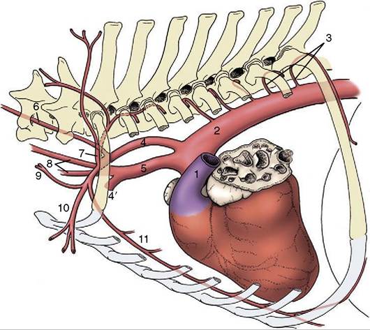
Figure 7-36 Branching of the aortic arch in the dog. (In this series of figures, not all arteries depicted are named.) 1, Pulmonary trunk; 2, aorta; 3, intercostal aa.; 4, left subclavian a.; 4', right subclavian a.; 5, brachiocephalic trunk; 6, vertebral a.; 7, costocervical trunk; 8, left and right common carotid aa.; 9, superficial cervical a.; 10, axillary a.; 11, internal thoracic a.
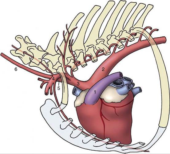
Figure 7-37 Branching of the aortic arch in the horse. The arteries to the head and neck and to the forelimbs originate from a short brachiocephalic trunk (3). 1, Pulmonary trunk; 2, aortic arch; 3, brachiocephalic trunk; 4, left subclavian a.; 5, bicarotid trunk; 6, left common carotid a.
follows the lateral attachment of the diaphragm. The cranial epigastric artery divides into superficial and deep branches; the latter follows the deep face of the rectus abdominis to an anastomosis with the caudal epigastric artery within the substance of this muscle.
The superficial branch passes to the superficial fascia, where it assists in the supply of the abdominal mammary glands.The superficial cervical artery (Figure 7—36/9), the fourth branch, arises from the subclavian opposite the origin of the internal thoracic. It supplies muscles of the ventral part of the neck, the cranial part of the shoulder, and the upper arm.
Aortic arch
Coronary aa.
Brachiocephalic trunk
Right subclavian a.
Vertebral a.
Costocervical trunk
Deep cervical a.
Internal thoracic a.
Ventral intercostal aa.
Cranial epigastric a.
Musculophrenic a.
Superficial cervical a.
Common carotid aa.
Left subclavian a. (its branches correspond to those of the right subclavian a.)
The Axillary Artery. The axillary artery (Figure 7-38/1), the magistral trunk of the forelimb, crosses the axilla to continue distally over the medial surface of the arm, caudal to the humerus. It changes its name again when level with the teres major tuberosity, where it becomes the brachial artery (Figure 7-38/6). The axillary gives external and lateral thoracic arteries to the chest wall and one important collateral branch to the limb, the subscapular artery (Figure 7-38/3). This runs dorsally along the caudal border of the scapula between the subscapularis and teres major. It supplies branches to the muscles of the shoulder.
The brachial artery (Figure 7-38/6) passes obliquely over the medial surface of the humerus to reach the craniomedial aspect of the elbow; it continues into the forearm where it shortly changes its name yet again, becoming the median artery. Its collateral branches include several to the muscles of the arm, principally the deep brachial (Figure 7-38/7) to the tricipital mass; toward the elbow it detaches collateral ulnar and superficial brachial arteries (Figure 7-38/8,9) that pass to the caudal and cranial aspects of the forearm, respectively. Branches of the superficial brachial run subcutaneously beside the cephalic vein and superficial branch of the radial nerve to reach the dorsum of the paw.
The transverse cubital artery (Figure 7-38/10) is detached just proximal to the elbow joint. A substantial branch, the common interosseous artery, originates from the main artery distal to the elbow.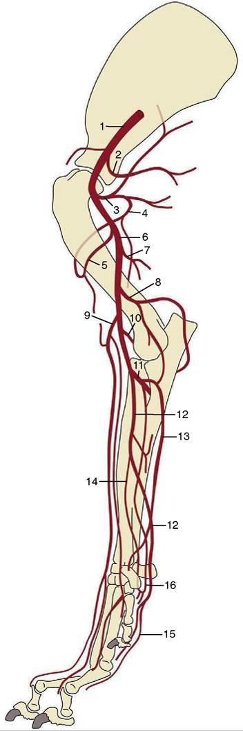
Figure 7-38 Arteries of the canine forelimb. 1, Axillary a.; 2, lateral thoracic a.; 3, subscapular a.; 4, caudal circumflex humeral a.; 5, cranial circumflex humeral a.; 6, brachial a.; 7, deep brachial a.; 8, collateral ulnar a.; 9, superficial brachial a.; 10, transverse cubital a.; 11, common interosseous a.; 12, median a.; 13, ulnar a.; 14, radial a.; 15, superficial palmar arch; 16, deep palmar arch.
The common interosseous artery (Figure 7-38/11) detaches the ulnar artery (Figure 7-38/13) for the digital and carpal flexors and the caudal interosseous artery, which runs between the radius and ulna to reach the palmar arches of the proximal metacarpus. A cranial interosseous penetrates the interosseous space to supply the dorsal muscles of the forearm.
The median artery (Figure 7-38/12) runs down the caudomedial aspect of the forearm in company with the median nerve and under protection of the flexor carpi radialis. It passes through the carpal canal to end by concurring with branches of the common interosseous in forming palmar arterial arches (Figure 7-38/15,16) from which the palmar aspect of the forepaw is supplied.
The paw receives its principal blood supply on its palmar aspect where (deep) palmar metacarpal and (more superficial) palmar common digital arteries run at the boundaries of the metacarpal bones before dividing at their distal ends into proper palmar digital arteries that follow the axial borders of the digits. The corresponding but narrower dorsal common and proper digital arteries follow a similar pattern.
Axillary a.
External thoracic a.
Lateral thoracic a.
Subscapular a.
Brachial a.
Deep brachial a.
Collateral ulnar a.
Superficial brachial a.
Cranial superficial antebrachial a.
Dorsal common digital aa.
Transverse cubital a.
Common interosseous a.
Ulnar a.
Cranial interosseous a.
Caudal interosseous a.
Superficial palmar arch
Palmar common digital aa.
Deep palmar arch
Palmar metacarpal aa.
Median a.
Radial a.
(The small arteries of the forepaw arise from anastomoses not listed.)
The Common Carotid Artery. The common carotid arteries arise separately in the dog (Figure 7-36/8) and by a short common (bicarotid) trunk in ungulates (Figure 7-37/5). Each crosses the ventrolateral face of the trachea (or esophagus on the left) in its ascent of the neck where it is accompanied by the vagosympathetic trunk. The artery ends by dividing above the
larynx into external and internal carotid arteries. The only significant collateral branches of the common carotid are detached close to its termination; they are the caudal and cranial thyroid arteries, of which the latter is the origin of the laryngeal and pharyngeal branches.
The external carotid artery is the larger of the terminal branches and appears as the direct continuation of the parent trunk (Figure 7-39/1,2). In the dog it shortly detaches the occipital artery, which branches from the internal carotid in some other species. The external carotid is continued as the maxillary artery (Figure 7-39/11); this distinction is rather arbitrarily determined by the origin of the superficial temporal artery.
The external carotid in this narrow sense forms a short dorsally convex arch resting on the pharynx and covered by the mandibular gland and digastricus. Its branches are the occipital, cranial laryngeal, ascending pharyngeal, lingual, facial, caudal auricular, parotid, and superficial temporal arteries.
The occipital artery (Figure 7-39/4) runs to the condyloid fossa where it divides into several branches that supply, among other structures, the middle and internal ear and the caudal meninges.
The largest branch, effectively the continuation of the stem, passes to the atlantal fossa to an anastomosis with the vertebral; it thus takes part in the supply to the brain (p. 312).The cranial laryngeal and ascending pharyngeal arteries (Figure 7-39/5,6) are the principal supplies to these organs (i.e., the larynx and pharynx). The large lingual artery (Figure 7-39/7) pursues a rostroventral course over the pharynx to enter the tongue between the genioglossus and hyoglossus muscles. It principally supplies the tongue, but collateral branches detached en route include one to the palatine tonsil that is of potential importance to the surgeon (p. 393).
The facial artery (Figure 7-39/8) arises near the angle of the jaw and runs within the intermandibular space before winding around the ventral border of the mandible where it is conveniently located for pulse taking in larger species; it then divides into various branches for the lips, lateral nose, and angle of the mouth. The relatively large caudal auricular artery (Figure 7-39/9) generously supplies the external ear and associated muscles. The parotid artery supplies the parotid gland.
The superficial temporal artery (Figure 7-39/10) winds onto the face and runs forward to supply the masseter. In the dog it branches to the upper and lower eyelids and dorsum of the nose. The position and firm support of one of the branches (transverse facial artery) suit it to pulse taking in larger species.
The maxillary artery (Figure 7-39/11) heads in the direction of the alar canal through which it passes to enter the pterygopalatine fossa. Before reaching the canal, its main branch is the inferior alveolar (Figure 7-39/12), which enters the mandible to supply the alveoli and teeth and, through mental branches that emerge from the bone, the lower lip and the chin region. Other maxillary branches pass to the tympanic cavity, muscles of mastication, and cranial meninges (the last passing through the oval foramen). No branches are detached from the stretch of artery within the canal, but a sheaf of diverging vessels comes off directly as it reaches the pterygopalatine fossa. The most important is the external ophthalmic artery (Figure 7-39/73) going to the contents of the orbit (p. 344). Others include the ethmoidal artery to the nasal cavity, the major and minor palatine arteries to the hard and soft palates, respectively, and the continuation (infraorbital artery) of the main trunk into the superior alveolar canal (Figure 7-39/74).

Figure 7-39 Arteries of the canine head. 1, Common carotid a.; 2, external carotid a.; 3, internal carotid a.; 4, occipital a.; 5, cranial laryngeal a.; 6, ascending pharyngeal a.; 7, lingual a.; 8, facial a.; 9, caudal auricular a.; 10, superficial temporal a.; 11, maxillary a.; 12, inferior alveolar a.; 13, external ophthalmic a.; 14, infraorbital a.
The internal carotid artery (Figure 7-39/3) enters the cranial cavity through the jugular foramen and carotid canal, taking a rather indirect course in the dog (p. 311). It divides within the cavity into divergent caudal and rostral branches that concur with their contralateral counterparts and with the basilar artery in forming the arterial circle from which the brain is supplied (p. 311). Common carotid a.
Caudal thyroid a.
Cranial thyroid a.
External carotid a.
Occipital a.
Cranial laryngeal a.
Ascending pharyngeal a.
Lingual a.
Facial a.
Caudal auricular a.
Parotid a.
Superficial temporal a.
Maxillary a.
Inferior alveolar a.
External ophthalmic a.
Ethmoidal a.
Palatine aa.
Infraorbital a.
Internal carotid a.
The Thoracic Aorta. The thoracic aorta runs cau- dally below the roof of the thorax to enter the abdomen by the aortic hiatus of the diaphragm. It continues as the abdominal aorta in company with the azygous vein and thoracic duct. The branches of the thoracic aorta are dorsal intercostal arteries (excepting those to the first few spaces), which arise variously and often by common trunks for the right and left vessels, and a broncho- esophageal artery, which is rather erratic in its origin.
Despite their names, which suggest rather restricted distribution within the intercostal spaces, the dorsal intercostal arteries detach substantial branches to the vertebral column and associated structures. They end by anastomosing with ventral intercostal arteries from the internal thoracic artery and its musculophrenic branch, thereby completing arterial loops within the spaces. The corresponding artery behind the last rib is known as the dorsal costoabdominal. The broncho- esophageal artery descends to the root of the lungs where it gives rise to bronchial branches for the tissues of the lungs and esophageal branches for much of the thoracic esophagus.
Thoracic aorta
Dorsal intercostal aa.
Bronchoesophageal a.
Bronchial branches Esophageal branches
Dorsal costoabdominal a.
The Abdominal Aorta. The abdominal aorta follows the roof of the abdomen, related to the caudal vena cava on its right and the psoas muscles on its left. Shortly after releasing the paired external iliac arteries, the abdominal aorta terminates in the dog below the last lumbar vertebra by branching off the internal iliac arteries and continues as the much smaller median sacral artery that extends into the tail (Figure 7-40/2,3,4). Along its course the abdominal aorta detaches both visceral and parietal branches.
The visceral arteries have been considered with the organs they supply. They comprise the unpaired celiac (p. 126), cranial mesenteric (p. 134), and caudal mesenteric (p. 134) arteries and the paired renal (p. 180) and testicular (p. 189 or ovarian [p. 203]) arteries. The unpaired vessels represent the arteries of the caudal foregut, midgut, and hindgut of the embryo (see Figure 3-65).
The collateral parietal branches begin with the caudal phrenic and cranial abdominal arteries, which share a common phrenicoabdominal origin in the dog. They also include the paired lumbar arteries to the tissues and structures of the back, the deep circumflex iliac to the flank, the external iliac artery to the hindlimb, and the internal iliac artery, which serves both pelvic viscera and pelvic walls.
Abdominal aorta
Phrenicoabdominal aa.
Lumbar aa.
Celiac a.
L. gastric a.
Hepatic a.
Hepatic branches
R. gastric a.
Gastroduodenal a.
Cranial pancreaticoduodenal a.
R. gastroepiploic a.
Splenic a.
Pancreatic branches
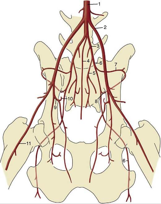
Figure 7-40 Termination of the canine abdominal aorta (ventral view). 1, Aorta; 2, external iliac a.; 3, internal iliac a.; 4, median sacral a.; 5, internal pudendal a.; 6, caudal gluteal a.; 7, iliolumbar a.; 8, cranial gluteal a.; 9, deep femoral a.; 10, pudendoepi- gastric trunk; 11, femoral a.
Short gastric aa.
L. gastroepiploic a.
Cranial mesenteric a.
Caudal pancreaticoduodenal a.
Jejunal aa.
Ileal aa.
Ileocolic a.
Middle colic a.
R. colic a.
Cecal aa.
Renal aa.
Testicular (ovarian) aa.
Caudal mesenteric a.
L. colic a.
Cranial rectal a.
Deep circumflex iliac aa.
External iliac aa.
Internal iliac aa.
Median sacral a.
Lumbar a. VI
Median caudal a.
It is worth drawing attention at this point to the existence of several pathways, established by anastomosis, that mitigate the effects of constriction or blockage of the aorta (e.g., by thrombosis, especially common in the cat). The collateral pathways include those formed along the spinal cord by anastomoses between successive lumbar arteries, those along the gut formed by connections between the principal visceral arteries, and those within the abdominal floor formed by the cranial and caudal epigastric arteries.
The External Iliac Artery. This is the principal artery of the hindlimb. It arises close to the termination of the aorta and runs obliquely over the abdominal roof to leave the abdomen by the vascular lacuna above the caudodorsal corner of the flank (Figure 7-41/5). It detaches one branch within the abdomen, the deep femoral artery (Figure 7-41/12), which is the common origin of the pudendoepigastric trunk and an important branch to the adductor muscles of the thigh. The short pudendoepigastric trunk (Figure 7-41/75) ends by giving rise to the caudal epigastric and external pudendal arter-
ies. The former divides in similar fashion to the cranial epigastric; the latter passes through the inguinal canal to supply structures in the groin, including the prepuce in the male and the caudal mammary glands (via the caudal superficial epigastric artery) in the bitch (see Figure 14-2).
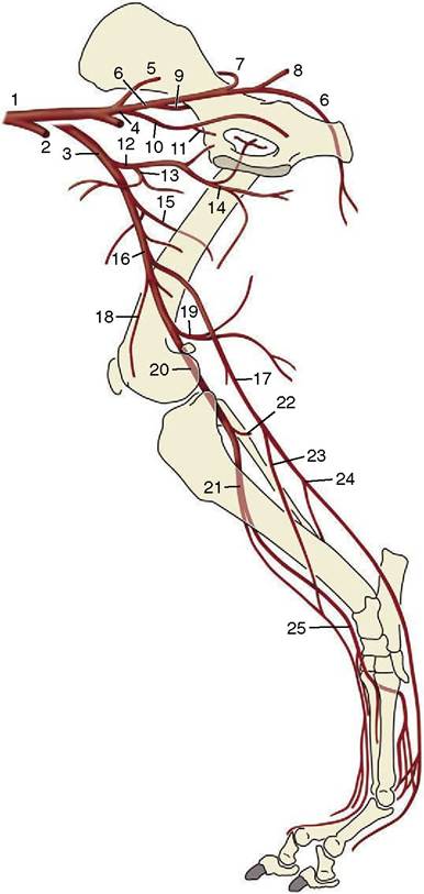
Figure 7-41 Arteries of the canine hindlimb. 1, Abdominal aorta; 2, left external iliac a.; 3, right external iliac a.; 4, left and right internal iliac aa.; 5, median sacral a.; 6, caudal gluteal a.; 7, cranial gluteal a.; 8, lateral caudal a.; 9, iliolumbar a.; 10, internal pudendal a.; 11, vaginal (prostatic) a.; 12, deep femoral a.; 13, pudendoepigastric trunk; 14, medial circumflex femoral a.; 15, lateral circumflex femoral a.; 16, femoral a.; 17, saphenous a.; 18, descending genicular a.; 19, distal caudal femoral a.; 20, popliteal a.; 21, cranial tibial a.; 22, caudal tibial a.; 23, cranial branch of the saphenous a.; 24, caudal branch of the saphenous a.; 25, dorsal pedal a.
The external iliac continues as the femoral artery (Figure 7-41Z16) on leaving the abdomen. Its first part has a superficial position in the femoral triangle— between the sartorius and pectineus, where it raises a visible ridge and is ideally located for pulse taking. It then burrows more deeply among the muscles to cross the medial surface of the femur to gain the caudal aspect of the thigh; it continues directly over the capsule of the stifle joint as the popliteal artery. The femoral artery has many branches, named and unnamed, to the muscles of the thigh but most do not require individual notice. One branch that does merit attention is the saphenous artery (Figure 7-41Z17), which is detached in midthigh. This is a more important vessel in carnivores than in the larger species; it descends over the medial aspect of the limb before dividing into cranial and caudal branches. The cranial branch (Figure 7-41Z25) supplies the dorsal crural muscles before crossing the dorsal aspect of the hock to continue as the dorsal common digital arteries. The caudal branch (Figure 7-41Z24) takes a deep course between the muscles of the caudal aspect of the leg (crus), which it supplies, crosses the caudal face of the hock, and terminates as the plantar common digital arteries, which are comparable to the corresponding forelimb arteries.
The popliteal (Figure 7-41Z20) divides into cranial and caudal tibial arteries. The cranial tibial artery (Figure 7-41Z21) passes through the interosseous space between the tibia and fibula to run distally with the deep peroneal nerve. It crosses the dorsal aspect of the hock (as the dorsal pedal artery; Figure 7-41Z25) and gives rise to the dorsal metatarsal arteries among other branches. One of these metatarsal arteries reinforces the caudal branch of the saphenous on the plantar aspect of the limb after passing between the second and third metatarsal bones. The caudal tibial artery (Figure 7-41Z22) is of little account in carnivores. The following list includes various muscular branches not mentioned in the text.
External iliac a.
Deep femoral a.
Pudendoepigastric trunk
Caudal epigastric a.
External pudendal a.
Femoral a.
Lateral circumflex femoral a.
Proximal, middle, and distal caudal femoral aa. Saphenous a.
Cranial branch
Dorsal common digital aa.
Caudal branch
Plantar common digital aa.
Popliteal a.
Cranial tibial a.
Dorsal pedal a.
Dorsal metatarsal aa. Plantar metatarsal aa.
Caudal tibial a.
The Internal Iliac Artery. This is the supply of the pelvic viscera and walls, including the overlying muscles of the gluteal region and those of the proximocaudal part of the thigh. The internal iliac artery continues caudoventrally from its origin, and in the dog it has a single branch, the umbilical artery (Figure 7-42/5), a rather unimportant vestige of the placental supply of the fetus (p. 255). The proximal part of the umbilical artery carries a little blood to the cranial part of the bladder; the distal part is transformed into the round ligament of the bladder within the lateral vesical fold.
The internal iliac artery terminates by dividing into the caudal gluteal and internal pudendal arteries. The parietal branch, the caudal gluteal artery (Figure 7-42/6), turns out of the pelvis with the sciatic nerve. This trunk, with its iliolumbar and cranial gluteal (Figure 7-42/7) branches, supplies the muscles about the lumbosacral junction and those of the gluteal and proximocaudal femoral regions; the structures of the last-named region include the proximal parts of the hamstring muscles in which the caudal gluteal terminates.
The second terminal branch is the internal pudendal artery (Figure 7-42/8) to the pelvic viscera (see also pp. 564 and 698). Its branches are differently named and disposed in the two sexes. The first branch is the prostatic artery in the male dog and the vaginal artery (Figure 7-42/9) in the female. The prostatic artery supplies the middle rectal artery to the penultimate part of the rectum and various branches to the caudal parts of the ureter and bladder, the prostate, and the first part of the urethra. The vaginal artery also supplies the rectum and urinary organs in addition to the uterus and vagina. Its cranial branch, the uterine artery, forms the caudal part of the arterial arcade within the broad ligament (p. 203).
The next artery, the urethral artery (Figure 7-42/10), is the same in both sexes. It supplies the caudal part of the pelvic urethra. The terminal branches of the internal pudendal are the ventral perineal artery and the artery of the penis or clitoris. The ventral perineal artery (Figure 7-42/11) supplies a caudal rectal artery to the last part of the rectum and branches to the scrotum (or labia of the vulva). The artery of the penis runs the length of the upper border of this organ to the region of the bulbus glandis; it becomes known as the dorsal artery of the penis after detachment of a branch to the penis bulb, which also supplies the corpus spongiosum and pars longa glandis, and a deep branch to the corpus cavernosum (p. 469 and Figure 15-20). The artery of the clitoris (Figure 7-42/12) is similar but on a less substantial scale.
Internal iliac a.
Umbilical a.
Caudal gluteal a.
Iliolumbar a.
Cranial gluteal a.
Internal pudendal a.
Prostatic (vaginal) a.
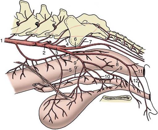
Figure 7-42 Arteries of the female pelvis, left lateral view (bitch). 1, Abdominal aorta; 2, external iliac a.; 3, internal iliac a.; 4, median sacral a.; 5, umbilical a.; 6, caudal gluteal a.; 7, cranial gluteal a.; 8, internal pudendal a.; 9, vaginal a.; 9', uterine a.; 10, urethral a. (frequently a branch of the vaginal a.); 11, ventral perineal a.; 12, a. of the clitoris.
A. of deferent duct (uterine a.)
Caudal vesicle a. Middle rectal a.
Urethral artery
Ventral perineal a.
Caudal rectal a.
Artery of penis (clitoris)
Artery of bulb
Deep artery Dorsal artery
The Systemic Veins
The systemic veins return blood to the heart through the cranial vena cava, caudal vena cava, and coronary sinus. The coronary sinus returns the bulk of the blood from the heart wall (p. 233); in ruminants and pigs it is joined by the left azygous vein. In the horse and the dog the equivalent (azygous) territory is drained by the right azygous.
The Cranial Vena Cava. The cranial vena cava is formed close to the entrance to the chest by the union of the external jugular and subclavian veins, which drain the head and neck and the forelimb, respectively. In the dog the subclavian and jugular veins of each side join in a common trunk, which then combines with its fellow; another arrangement is the union of the two jugulars in a single bijugular trunk, which is then joined by the subclavian veins. The cranial vena cava runs through the cranial mediastinum, ventral and to the right of the trachea, and is related to the brachiocephalic trunk (dorsally at its origin, later at its left face). It is joined by various tributaries broadly corresponding to branches of the subclavian artery and by the larger right azygous vein toward its termination (Figure 7-43/5)—unless this makes separate entry to the right atrium as in the horse.
The azygous vein (Figure 7-43/5) is formed by the union of the first lumbar veins and passes through the aortic hiatus into the chest where it is reinforced by intercostal veins from the caudal and middle intercostal spaces. Right and left veins are present in the embryo, but the pattern is later commonly simplified: the main trunk is the right azygous vein in horses and dogs and the left one in ruminants and pigs—unless, as is usual in ruminants, both remain of some size. The right azygous vein arches ventrally, passing in front of the root of the right lung to reach the terminal part of the cranial vena cava or the adjacent part of the right atrium (horse). The left vein arches in front of the root of the left lung and must then run caudally, over the left atrium, to reach its confluence with the coronary sinus (Figure 7-9, A/12). The cranial intercostal veins that do not drain into this system join various tributaries of the subclavian or go directly to the cranial vena cava. The special importance of the azygous system in draining the plexus within the vertebral canal is considered elsewhere (p. 314).
The subclavian vein generally corresponds to the subclavian artery, and most tributaries in the upper part of the limb are satellite to arterial branches. The pattern is different in the distal part of the limb where important unaccompanied superficial veins are present. Although these are connected with the deeper veins at various levels, they also continue into the cephalic vein (Figure 7^43/15), which runs between the pectoral and brachiocephalic muscles in the arm to join the external jugular vein in the lower part of the neck.
Two pairs of jugular veins exist within the neck. The deep internal jugular (Figure 7-43/5) runs with the common carotid artery within the visceral space of the neck; however, except in the dog and cat, it is very much reduced in size or even absent in postnatal animals. Even in the dog and cat it is of minor importance. The external jugular vein (Figure 7-43/6) is formed near the angle of the jaw by the union of linguofacial and maxillary veins. Its course through the neck occupies a (jugular) groove between the brachiocephalicus dorsally and the sternocephalicus ventrally in the larger species; in the dog it lies on the sternocephalicus. It is easily raised for intravenous injection and blood sampling, and in the larger species it is the first choice for these procedures. The territories of its linguofacial and maxillary tributaries show considerable overlap and some species variation; the former vein is in general the principal drainage of the more superficial and more rostral structures of the head, the latter of those deeper and more caudal, including the contents of the cranial cavity (see Figure 11-44).
The Caudal Vena Cava. The caudal vena cava is formed on the roof of the abdomen, near the pelvic inlet, by the union of right and left common iliac veins, each formed in its turn by the union of an internal iliac vein, which drains the pelvic walls and much of the contents of the pelvic cavity, and an external iliac vein, which drains the hindlimb (Figure 7-43/25,51). The external iliac vein and the bulk of its tributaries are satellite to arteries. The independent medial and lateral saphenous veins of the leg (Figure 7-43/55,57) drain the superficial veins of the foot.
In its intraabdominal course the caudal vena cava is joined by additional tributaries draining the abdominal roof, including large renal veins, before it dips ventrally to tunnel through the liver and subsequently the diaphragm at the caval foramen. It enters the thoracic cavity at a relatively ventral level and pursues a course within the free edge of the plica venae cavae between the caudal and accessory lobes of the right lung (see Figure 4-20, B/9). It joins the right atrium dorsal to the inlet of the coronary sinus.
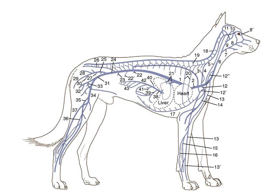
Figure 7-43 Schematic representation of the venous system (dog). 1, Caudal vena cava; 2, cranial vena cava; 3, azygous v.; 4, vertebral v.; 5, internal jugular v.; 6, external jugular v.; 7, linguofacial v.; 8, facial v.; 8’, angularis oculi v.; 9, maxillary v.; 10, superficial temporal v.; 11, dorsal sagittal sinus; 12, subclavian v.; 12’, axillobrachial v.; 12", omobrachial v.; 13, cephalic v.; 13', accessory cephalic v.; 14, brachial v.; 15, radial v.; 16, ulnar v.; 17, internal thoracic v.; 18, vertebral venous plexus; 19, intervertebral v.; 20, intercostal vv.; 21, hepatic vv.; 22, renal v.; 22', testicular or ovarian v.; 23, deep circumflex iliac v.; 24, common iliac v.; 25, right internal iliac v.; 26, median sacral v.; 27, prostatic or vaginal v.; 28, lateral caudal v.; 29, caudal gluteal v.; 30, internal pudendal v; 31, right external iliac v.; 32, deep femoral v.; 33, pudendoepigastric trunk; 34, femoral v.; 35, medial saphenous v.; 36, cranial tibial v.; 37, lateral saphenous v.; 38, portal v.; 39, gastroduodenal v.; 40, splenic v.; 41, caudal mesenteric v.; 42, cranial mesenteric v.; 43, jejunal vv.
In its intrahepatic course the caudal vena cava receives the hepatic veins, which drain the liver (Figure 7-43/21).
The portal vein drains the spleen, the intraabdominal digestive organs, the caudal part of the thoracic esophagus, and the bulk of the rectum (Figure 7-43/38 and Figure 7-44). It is formed variously from three main tributaries (see Figure 3-50/2,4,5). The splenic tributary corresponds to the celiac artery (excluding its hepatic branches) and therefore drains the last part of the esophagus, the stomach, parts of the duodenum and pancreas, and the spleen. The cranial and caudal mesenteric veins drain the territories of the like-named arteries and usually join in a common trunk before combining with the splenic.
The last part of the rectum and the anal region differ from the remainder of the gut in draining toward the internal iliac vein. The veins of this part form one of the portosystemic connections that provide alternative (although not very capacious) outlets from the portal drainage territory that are used when the intrahepatic circulation is impaired, as, for example, by cirrhosis (hepatic fibrosis).