THE TEMPOROMANDIBULAR JOINT
The articular surfaces of the temporomandibular joint are nearly congruent. The transverse cylinder provided
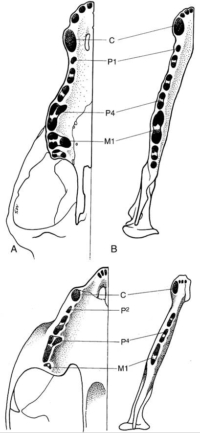
Figure 11-24 The tooth sockets in canine (top) and feline (bottom) upper (A) and lower (B) jaws to show the number and disposition of the roots.
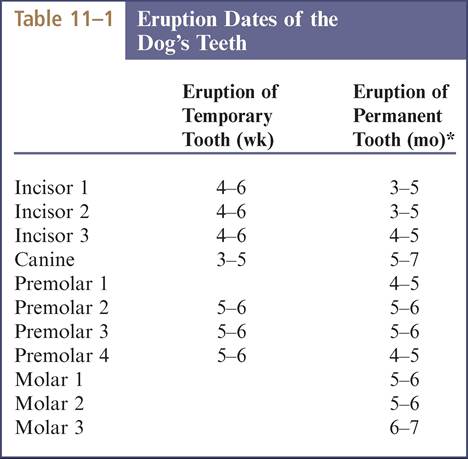
*Permanent teeth erupt slightly earlier in large breeds. Modified from Schummer A, Nickel R, Sack WO: The viscera of the domestic mammals, ed 2, New York, 1979, SpringerVerlag; and Evans HE: Miller's anatomy of the dog, ed 3, Philadelphia, 1993, Saunders.
by the mandible fits within a trough on the undersurface of the zygomatic process of the temporal bone (Figures 11-23, 11-28, and 11-29). The trough is enlarged cau- dally by a prominent retroarticular process that securely cups the cylinder and prevents its luxation in a caudal direction. In keeping with the congruence of the joint, the articular disk is thin. The joint capsule is strengthened by a lateral ligament.
Movement of the mandible is almost exclusively of a hinge nature; only slight protrusion is possible when the mouth is fully open. Lateral movement may be produced by trauma and occasionally is so severe that the
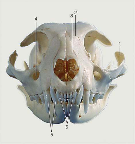
Figure 11-25 Feline skull, rostral view. 1, Zygomatic arch; 2, frontal bone; 3, nasal bones; 4, infraorbital foramen; 5, upper and lower canine teeth; 6, upper and lower incisors, in incisive bones and mandible, respectively.
From Schummer A, Nickel R, Sack WO: The viscera of the domestic mammals, ed 2, New York, 1979, SpringerVerlag.
coronoid process engages the zygomatic arch, locking the jaws in the depressed position.
The joint lies under cover of the caudal part of the masseter, where the dorsal buccal branch of the facial nerve crosses the border of the muscle. It is rostral to the parotid gland.
The masticatory muscles have been sufficiently described (p. 113).