PAROTID GLAND
The parotid gland (see Figure 11-6) is roughly triangular, relatively thin, and molded around the proximal portion of the auricular cartilage, against which it can be rolled on palpation.
It occupies a depression between the masseter, the wing of the atlas, and the auricular cartilage. Ventral to the cartilage, it is related medially to the facial nerve and maxillary vein and more rostrally to the parotid lymph node and temporomandibular joint. The parotid duct leaves the cranial aspect of the gland and continues over the lateral aspect of the masseter muscle between the buccal branches of the facial nerve. The duct opens into the vestibule at a small parotid papilla opposite the caudal part of the upper fourth premolar tooth, approximately 5 mm from the margin of the gum. The duct makes a right-angle bend just before opening at the papilla; cannulation is made easier by grasping the mucosa just caudal to the opening and pulling it rostrally to straighten the bend.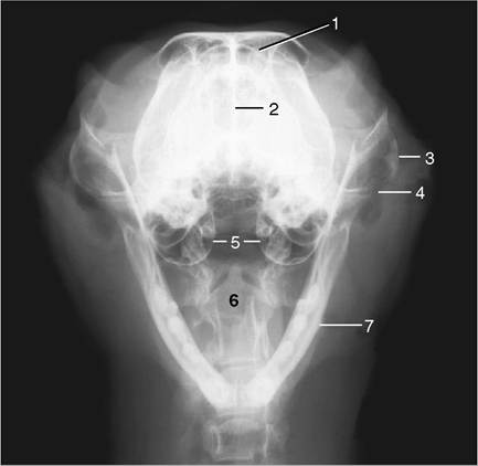
Figure 11-26 Rostrocaudal open-mouth radiograph of a feline head. 1, Frontal sinus; 2, nasal septum; 3, zygomatic arch; 4, temporomandibular joint; 5, tympanic bullae; 6, axis with dens; 7, mandible.
More on the topic PAROTID GLAND:
-
Veterinarian -