The term mouth (os, genitive form oris) designates the cavity, its walls, and also the accessory structures that project (teeth, tongue) and drain (salivary glands) into it.
The mouth has as its main functions the prehension, mastication, and insalivation of food. It may also play a role in aggression and defense, whereas in ourselves it is important in the formulation of the sounds of speech.
In most species it functions as an airway when flow through the nose is impaired.The mouth (oral cavity) is entered between the lips and continues into the pharynx (Fig. 3.3) through a caudal narrowing at the level of the palatoglossal arches (see later). It is divided by the teeth and margins of the jaws into an outer vestibule, bounded by the lips and cheeks externally and the central oral cavity proper. When the mouth is closed, these divisions communicate through gaps behind and between the teeth. The vestibule extends caudally toward the ramus of the mandible and the masseter muscle. The proportion of its walls formed by the lips varies with feeding habits; a wide gape is necessary in species that feed greedily or use their teeth to seize prey or in fighting, whereas a smaller opening suffices in most herbivores and rodents.
Diet and feeding habits also determine the form of the lips (labia oris). In some species, such as the horse, the lips are sensitive and mobile to accomplish the task of food collection and its introduction into the mouth. When other parts are more important in prehension the lips can be less mobile and smaller (e.g., cat) or thickened and insensitive (e.g., ox). The lips of the dog are extensive but thin and can be posed to show aggressive intent or submission. In newborn animals the lips form the seal about the teat that is necessary for successful sucking. The mimetic muscles that encircle the mouth and raise, depress, and retract the lips are supplied by the facial nerve.
The lips are composed of skin, an intermediate layer of muscle, tendon, and glands, and the oral mucosa. The skin and mucosa usually meet along the margin of the lips, though the boundary can be displaced in either direction.
Small salivary glands are scattered among the muscle bundles below the mucosa, especially toward the angles (commissures) where the two lips meet.Compared with the upper lip, the lower lip is usually unremarkable. In the dog it is rather loose but fastened to the lower jaw at the level of the canine tooth and has a thin, serrated margin. The upper lip sometimes has a median naked area that continues with the modified skin around the nostrils. The extensive moist and glandular nasolabial plate of the ox and the rostral disk of the pig are good examples of this. The area of modified skin, referred to by dog breeders as "nose leather," is often much narrower and may be divided by a median groove (philtrum) (see Fig. 3.3). In the human and in the horse a hairy integument extends across the entire upper lip.
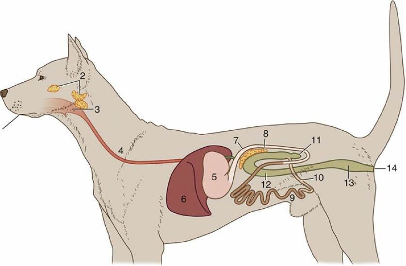
FIG. 3.1 Schematic representation of the digestive apparatus in the dog. 1, Mouth; 2, salivary glands; 3, pharynx; 4, esophagus; 5, stomach; 6, liver; 7, duodenum; 8, pancreas; 9, jejunum; 10, ileum; 11, cecum;
12, colon; 13, rectum; 14, anus.
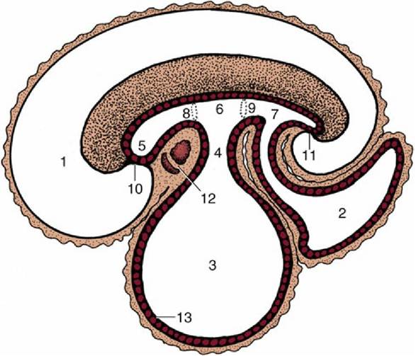
FIG. 3.2 Sagittal section of an early embryo. Part of the yolk sac is taken into the body in the folding process. 1, Amniotic cavity; 2, allantoic cavity; 3, yolk sac; 4, stalk of yolk sac; 5, foregut; 6, midgut; 7, hindgut; 8, cranial intestinal portal; 9, caudal intestinal portal; 10, oral plate; 11, cloacal plate; 12, heart and pericardial cavity; 13, endoderm.
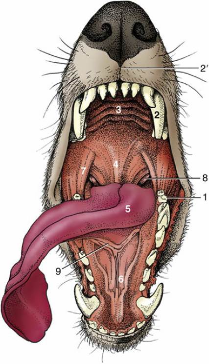
FIG. 3.3 General view of the oral cavity of the dog. 1, Vestibule; 2, canine tooth; 2', philtrum; 3, hard palate; 4, soft palate; 5, tongue; 6, sublingual caruncle; 7, palatoglossal arch; 8, palatine tonsil; 9, frenulum.
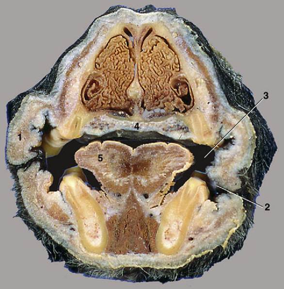
FIG.
3.4 Transverse section of the head of the dog at the level of P2. 1, Cheek (with buccal folds); 2, vestibule; 3, oral cavity proper; 4, hard palate (with venous plexus); 5, tongue.The cheeks (buccae), which are most capacious in herbivores, are structurally similar to the lips. The principal support is the buccinator muscle, which functions to return to the central cavity any food that has escaped into the vestibule. Certain rodents and monkeys have diverticula of the oral vestibule (cheek pouches) to store rapidly harvested food for later mastication. The large cheek pouches in hamsters reach well into the thorax and have their own supporting musculature. There are additional salivary glands, sometimes aggregated in quite large masses: the zygomatic gland of the dog (see Fig. 3.12/8) that are concealed below the zygomatic arch. The buccal mucosa tends to be tightly anchored in some places such that it is sufficiently loose to allow the occasional maximal opening of the mouth while avoiding large folds that would get injured from the teeth (Fig. 3.4). In ruminants, whose food may be dry and rough, large, closely spaced, pointed papillae provide protection (see Fig. 3.7). A small papilla (in h easily found with the tongue tip) carries the opening of the duct of the parotid gland.
The cavity within the dental arcades—the mouth cavity proper—is roofed by the palate; bounded laterally by the teeth, gums, and margins of the jaws; and floored by the tongue and the small area of mucosa left uncovered by the tongue. Most of the walls are rigid, and when the mouth is closed, the size of the cavity can be altered only by raising or lowering the tongue and floor.
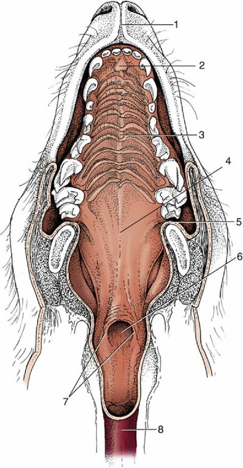
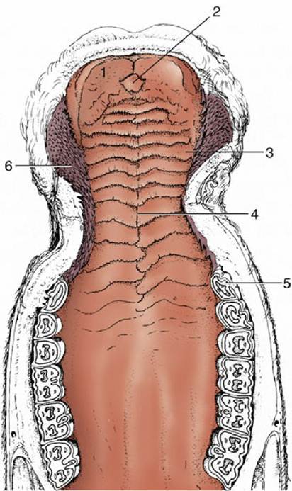
FIG. 3.5 The hard and soft palate of the dog. 1, Philtrum; 2, incisive papilla; 3, hard palate with rugae; 4, soft palate; 5, palatoglossal arch; 6, intrapharyngeal ostium; 7, palatopharyngeal arches; 8, esophagus.
The larger, rostral part of the roof is based on a bony shelf formed of the palatine processes of the incisive, maxillary, and palatine bones and is known as the hard palate (palatum durum). This structure is continued caudally, without external demarcation, by the soft palate, in which a connective tissue aponeurosis replaces the bone.
The hard palate is usually flat (though vaulted in humans) and is covered by a thick mucosa fashioned into a series of more or less transverse ridges (rugae), which may guide the food backward (Fig. 3.5). In general, these ridges are most prominent and their covering epithelium most heavily keratinized in herbivores. A small median swelling, the incisive papilla, is commonly found behind the incisor teeth, flanked by the orifices of small (incisive) ducts that perforate the palate. These ducts branch and lead to the nasal cavity and to the vomeronasal organ (Fig. 3.6). They convey small amounts of the fluid from the mouth for appraisal by the olfactory mucosa of the vomeronasal organ (p. 337).
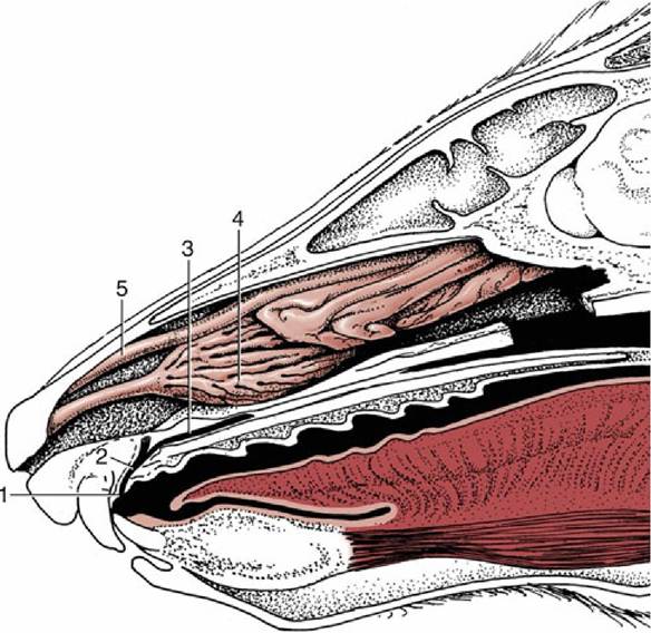
FIG. 3.6 Paramedian section of the rostral part of the head of the dog. The plane of section fails to demonstrate the opening of the incisive duct into the nasal cavity. 1, Incisive papilla; 2, incisive duct; 3, vomeronasal organ; 4, ventral nasal concha; 5, dorsal nasal concha.
FIG. 3.7
The hard palate of a cow. 1, Dental pad; 2, incisive papilla; 3, rugae of hard palate; 4, palatine
raphe; 5, P2; 6, buccal papillae.
A striking peculiarity in ruminants is the dental pad, a tough but yielding cushion in the position generally occupied by upper incisor teeth (lacking in these animals). The dental pad acts as a counterpart to the lower incisors in grazing (Fig. 3.7). A dense, richly vascularized tissue beneath the palatine epithelium functions both as the lamina propria of the mucosa and as the periosteum of the bone, attaching so tightly that not even the most vigorous mastication shifts it. Peripherally, the hard palate blends with the gums, the rather insensitive mucosa along the alveolar margins of the jaws.
The soft palate is described with the pharynx (p. 109).