The Tongue
The tongue (lingua) occupies the greater part of the oral cavity but also extends into the oropharynx (Fig. 3.8). It has an attached root and body and a free apex. The highly muscular construction makes the tongue capable of both vigorous and precise movements, as in prehension, lapping, grooming, and manipulating the food within the mouth on the one hand and speech articulation on the other.
The mobility is achieved by restriction of the attachments to the more caudal part, which leaves the apex free to move both within and beyond the mouth. The attachment of the root is to the hyoid bone, and that of the body is to the symphyseal region of the mandible. The tongue is slung between the lower jaws by paired mylohyoideus muscles that originate from the medial aspect of the mandible and meet in a median raphe (see Fig. 3.21/4). Dogs also use their tongues for heat loss by panting, a process facilitated by the very generous supply of blood and the numerous arteriovenous anastomoses (p. 226).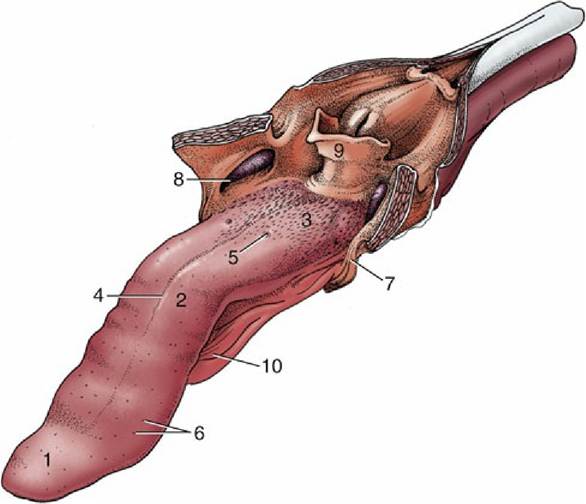
FIG. 3.8 The tongue of the dog. The soft palate and the esophagus are sectioned in the median plane. 1, Apex; 2, body; 3, root, forming floor of oropharynx; 4, median groove; 5, vallate papilla; 6, fungiform papillae; 7, palatoglossal arch; 8, palatine tonsil in tonsillar fossa; 9, epiglottis; 10, frenulum.
In general shape, the tongue corresponds to the oral cavity. The apex is dorsoventrally compressed, the succeeding middle portion is somewhat triangular in section (being joined to the oral floor by a mucosal fold or frenulum), and the root is uniformly wide to allow entry to the muscles passing forward from the hyoid bone. Mucosal reflections (palatoglossal arches; Fig. 3.8/7) also pass from each side of the root to join the soft palate and demarcate the boundaries of the mouth.
The mucosa is tough and tightly adherent where repeated contact with abrasive food occurs but looser and less heavily keratinized where a softer diet or a more protected position allows. Much of the surface is covered by a variety of papillae. Some are soft and threadlike (filiform), scattered widely over the human tongue to provide additional protection, but the harsh conical papillae make the cat's tongue essentially function as a rasp. Other papillae carry taste buds and have a more restricted distribution, characteristic for each species (Fig. 3.9): their names—fungiform, foliate, and vallate papillae—give good indications of their shapes. A few small salivary glands lie below the epithelium.
The bulk of the tongue consists of muscle, usually divided into intrinsic and extrinsic groups. Four pairs of extrinsic muscles exist (Fig. 3.10). One, the geniohyoideus, lies somewhat apart and below the tongue and passes from the incisive part of the mandible to the body of the hyoid bone. It is able to draw the hyoid and thus the tongue forward. The genioglossus arises more dorsally than the geniohyoideus and first runs back below the floor of the mouth before dividing into bundles that fan upward in the sagittal plane. Those bundles going to the apex of the tongue retract this part while those to the root draw the whole tongue forward. The middle group passes toward the upper surface (dorsum), which it may depress. The other two muscles arise from the hyoid apparatus. The hyoglossus takes origin from the basihyoid and runs forward, lateral to the genioglossus; the styloglossus takes origin from the stylohyoid but farther to the side. Both draw the tongue back but in rather different fashions; the styloglossus also tends to elevate it. The bundles of intrinsic muscle run longitudinally, transversely, and vertically (see Fig. 4.2). Simultaneous contraction of the transverse and vertical bundles stiffens the tongue.
The muscle bundles are interspersed with considerable amounts of fat, which is an arrangement that imparts a unique consistency and flavor to the cooked tongue.
This fat is very resistant to mobilization in starvation.In the dog, alone among the domestic species, the ventral part of the tongue contains a prominent fibrous condensation, the lyssa, easily recognized on palpation. A fibrous septum that extends from this is responsible for the conspicuous median groove on the upper surface.
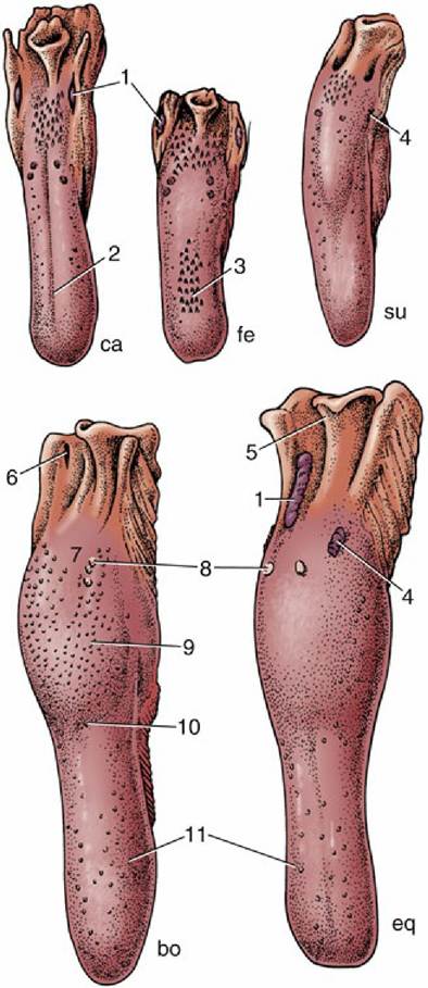
FIG. 3.9 Dorsal view of the tongue and epiglottis of the dog (ca), cat (fe), pig (su), cattle (bo), and horse (eq). 1, Palatine tonsil; 2, median groove; 3, filiform papillae; 4, foliate papillae; 5, epiglottis; 6, tonsillar sinus; 7, root of tongue; 8, vallate papillae; 9, torus linguae; 10, fossa linguae; 11, fungiform papillae.
The innervation accurately reflects the origin of the tongue as an unpaired swelling of the pharyngeal floor (see Fig. 3.58C) that is later extended by contributions from the ventral parts of the adjacent pharyngeal (branchial) arches. The mucosa retains a sensory innervation from the corresponding arch nerves. The lingual branch of the mandibular nerve is responsible for general sensation over the rostral two thirds of the tongue; the chorda tympani, a branch of the facial nerve, is responsible for the special sensation of taste in the same area. Both general and special sensations of the root region are the responsibility of the glossopharyngeal and, to a small extent, the vagus nerves.

FIG. 3.10 Muscles of the tongue and pharynx of the dog. 1, Geniohyoideus; 2, mylohyoideus; 3, genioglossus; 4, styloglossus; 5, hyoglossus; 6, sternohyoideus; 7, sternothyroideus; 8 and 9, hyopharyngeus (two parts); 10, thyropharyngeus; 10', cricopharyngeus; 11, thyrohyoideus; 12, cricothyroideus.
The extrinsic and intrinsic muscles are all supplied by the hypoglossal nerve, although it is probable that the sensory fibers emanating from spindles and other receptors in these muscles travel mainly in the lingual nerve. The mylohyoideus muscle is supplied by the mandibular nerve and plays an important part in initiating swallowing.
Relatively little of the floor of the mouth is left accessible rostral and lateral to the attachments of the tongue. The largest free area lies ventral to the apex, behind the incisor teeth. The mucosa here covers the incisive part of the mandible directly, but elsewhere it lies on muscle and the floor yields under pressure. The most prominent features are fleshy protuberances or caruncles behind the central incisors, and located in them are the common openings of the mandibular and major sublingual salivary ducts (see Fig. 3.3). In some species, much smaller serial elevations to each side of the frenulum mark the openings of the lesser ducts of the sublingual gland.