THE TRACHEA
The trachea and bronchi form a continuous system of tubes conducting air between the larynx and the smaller passages (bronchioli) in the lungs. They have a very similar construction and together are sometimes termed the tracheobronchial tree.
The trachea leads from the larynx through the visceral space of the neck, enters the mediastinum at the thoracic inlet, and continues to its terminal bifurcation above the heart. The two chief bronchi diverge from the line of the trachea to enter the corresponding lungs at their roots. In ruminants and pigs a separate tracheal bronchus arises proximal to the tracheal bifurcation and separately aerates the cranial lobe of the right lung. The cervical part of the trachea maintains a more or less median position, although its relationship to the esophagus alters at different levels and in different postures of the head and neck (see Figures 3-29 and 4-16/7). Other relations in the neck include the ventral strap muscles of the neck and the carotid sheath and its contents; the common carotid artery commences ven- trolaterally but gradually climbs to a dorsolateral position where the trachea originates from the larynx.
The thoracic part of the trachea is deflected slightly to the right where it crosses the aortic arch. It is related ventrally to the cranial vena cava, to the arteries arising from the aortic arch, and to various tributaries and branches of these vessels; it is related dorsally to the esophagus and related variously to mediastinal lymph nodes. In young subjects it is related to the thymus. The bifurcation lies in the region of the fourth to sixth intercostal spaces but varies with the species and with the respiratory phase.
The chief bronchi very quickly enter the lungs (Figure 4-17), in which they ramify according to a pattern described later (p. 162).
The wall of the trachea is composed of an inner mucosa, a fibrocartilaginous middle layer, and an adventitia (in the neck) or serosa (in the thorax) (Figure 4-18).
The mucosa, which continues that lining the infraglottic part of the larynx, may show slight longitudinal folding when the lumen is narrowed. It contains both unicellular and multicellular mucous glands that produce a protective covering of mucus that is continuously moved toward the larynx by the ciliary action of the epithelium. This mucus eventually reaches the pharynx and is swallowed without being noticed. Excessive mucus accumulations may irritate the mucosa, stimulating coughing to clear the airway. The fibrocartilaginous coat is composed of numerous strips of cartilage that are bent to form “rings” that are incomplete dorsally where the ends may fail to meet or may overlap.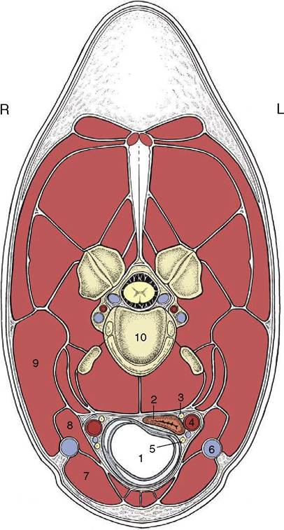
Figure 4-16 Transverse section of the neck (horse) at the level of the fourth cervical vertebra. 1, Trachea; 2, esophagus; 3, vagosympathetic trunk; 4, common carotid artery; 5, caudal (recurrent) laryngeal nerve; 6, external jugular vein; 7, sterno- cephalicus; 8, omohyoideus; 9, brachiocephalicus; 10, body of the fourth cervical vertebra.
The edges of the strips are connected to each other by sheets of rather elastic connective tissue continuous with the perichondrium. The ends are joined by the smooth tracheal muscle (Figure 4-18∕√), which bridges the gap within the “ring” in most species but is placed externally in the dog and the cat.
The construction of the trachea prevents it from collapsing and allows it to make the necessary adjustment in length when the neck is extended and also when the diaphragm contracts. It is attached to the diaphragm indirectly by the pulmonary ligaments and mediastinal connective tissue and also, more effectively, by the nega-
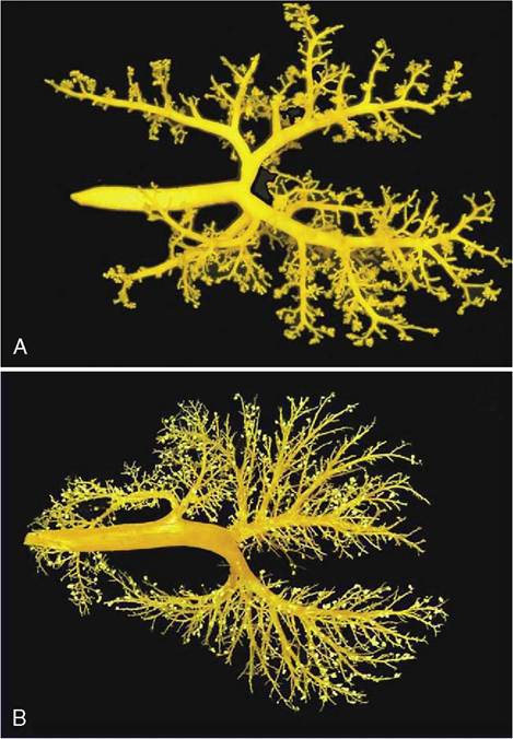
Figure 4-17 Dorsal views of corrosion casts of the bronchial tree and lungs of the cat (A) and calf (B).
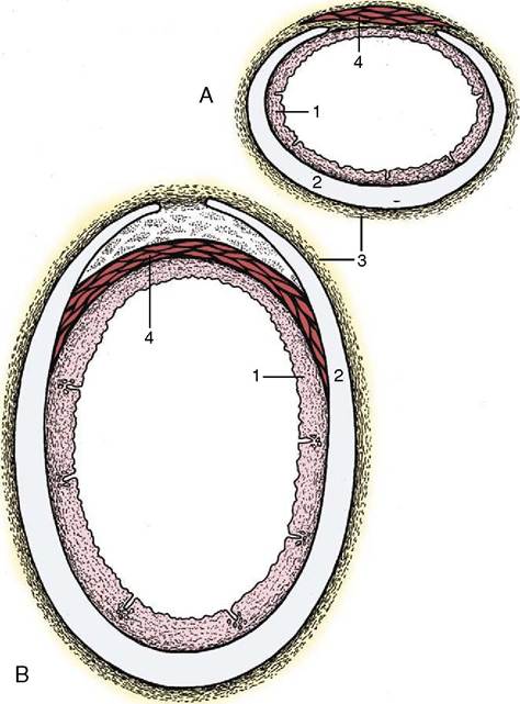
Figure 4-18 Transverse sections of the canine (A) and bovine (B) trachea. 1, Mucous membrane; 2, tracheal cartilage; 3, adventitia; 4, tracheal muscle (external in dogs, internal in cattle).
tive intrapleural pressure that couples the lungs to the chest wall, including the diaphragm. Variations in diameter are regulated by the tracheal muscle. In addition to these functional changes, there are permanent species and regional variations in the cross-sectional form and area of the trachea.
The structure of the larger bronchi is identical to that of the trachea if allowance is made for the mergence of their outer surfaces with the peribronchial connective tissue (and through this with the stroma of the lung). On the smaller bronchi the cartilage rings are gradually replaced by irregular plaques, and it is the shedding of the last of these that defines the bronchobronchiolar transition.
Variations in the diameter of the bronchi and bronchioli are relatively greater and more significant than those of the trachea.
Before proceeding, one may need to reread the section on the shape and function of the thoracic cavity (p. 52).