THE PLEURA
Each lung is invested by a serous membrane, the pleura, which also lines the corresponding “half” of the thoracic cavity. Thus, two pleural membranes exist, each arranged as a closed invaginated sac.
The space between the right and left sacs forms the mediastinum, a more or less median partition in the thorax within which the heart and other thoracic organs are situated (Figure 4-19/7).The part of the pleura that clothes the lung directly is known as the visceral or pulmonary pleura (Figure 4-19∕√). It is reflected around, and also behind, the root of the lung to become continuous with the mediastinal pleura which, in turn, is continuous with the costal and diaphragmatic pleura; these last three parts are together termed the parietal pleura.
In the healthy animal the pleural cavity is a potential rather than an actual space, and it contains only a small
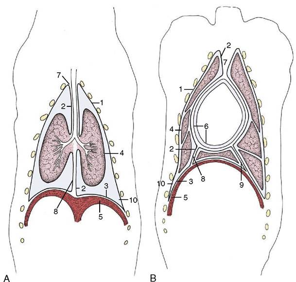
Figure 4-19 Schematic dorsal sections of the pleural cavities (dog); at the level of the tracheal bifurcation (A) and at the level of the heart (B). 1-3, Parietal pleura, later subdivided; 1, costal pleura; 2, mediastinal pleura; 3, diaphragmatic pleura; 4, visceral pleura; 5, diaphragm; 6, parietal and visceral pericardium; 7, cranial mediastinum; 8, caudal mediastinum; 9, plica venae cavae; 10, costodiaphragmatic recess.
amount (a few milliliters) of serous fluid, which is thinly spread over the pleural surface and facilitates the smooth movement of the lung against the chest wall and of one lung lobe against another. The pressure within the pleural cavity, which is about -5 cm H2O in the neutral resting position of the chest, represents the difference between the forces that tend to recoil the lung and those that tend to expand the chest. The pressure is not uniform throughout the pleural cavity, and in addition to the expected dorsoventral gradient, local and partly unexplained differences exist; these variations in intrapleural pressure account for regional differences in the expansion and aeration of the lungs.
The prevailing negative pressure explains why a surgical or traumatic opening in the chest wall causes an inrush of air into the pleural cavity, collapsing the lung and producing the condition known as pneumothorax.The pleural sac is always more extensive than the lung, and in certain regions, facing surfaces of parietal pleura are directly applied to each other. The most important example of such an arrangement is found caudal to the basal border of the lung, where the peripheral part of the diaphragmatic pleura rests against the costal pleura lining the chest wall (the costodiaphragmatic recess; Figure 4-19/70). Although the extent of the recess varies with the phase of respiration, it remains considerable even in full inspiration, and the potential of this portion of the pleural sac is therefore never realized (see Figure 4-22/6). A similar but smaller costomediastinal recess is present ventral to the lung (Figure 4-20/72).
Cranially, the costal and mediastinal portions of the pleura come together to form a dome, the cupula pleurae, which may extend in front of the first rib, where it is obviously vulnerable to injury (Figure 4-2W'). The mediastinum is not symmetrical but is deflected to the left at certain levels. The important deflection of the caudal mediastinum is produced by the greater size of the base of the right lung.
A special fold (plica venae cavae) of the pleura of the right sac extends between the diaphragm and pericardium and carries the caudal vena cava in its free dorsal border (Figure 4-20/3,9). This triangular partition helps define a recess into which the accessory lobe of the right lung fits (Figure 4-21).
Considerable practical significance attaches to the strength of the mediastinum, which varies much between species. In some, for example, the ruminants, the
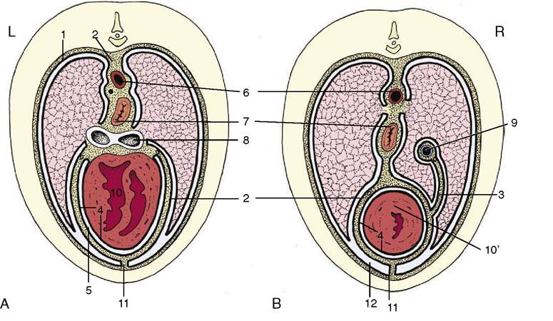
Figure 4-20 Schematic transverse section of the thorax at the level of the heart (A) and at the transition of heart to caudal mediastinum (B). 1, Costal pleura; 2, mediastinal pleura; 3, plica venae cavae; 4, parietal and visceral pericardium; 5, pericardial space; 6, aorta; 7, esophagus; 8, tracheal bifurcation; 9, caudal vena cava; 10, heart; 10', apex of heart; 11, sternopericardial ligament; 12, costomediastinal recess.
mediastinum is thick and able to withstand a considerable pressure difference between the two pleural cavities; consequently, collapse of one lung may be tolerated. In others, for example, the dog, cat, and horse, it is very delicate and ruptures readily. Indeed the horse is among those species in which the mediastinum of the dead specimen always presents numerous small openings that place the right and left pleural cavities in communication.