THE UDDER
The mammary glands are consolidated in a rather small udder situated below the caudal part of the abdominal floor and cranial part of the pelvis and concealed from casual inspection by the thigh (Figure 22-25).
The form and size of the udder vary with the present state and previous history of the mare; the udder is very small in young virgin animals. A prominent external groove indicates its formation from right and left halves; each half has the form of a laterally compressed cone and, though carrying a single teat, is composed of two (occasionally three) separate duct systems.The skin over the udder is thin, strongly pigmented, and sparsely haired; it is supplied with many sweat and sebaceous glands and usually glistens. The teat is small and cylindrical, except in the lactating mare, in which it is both larger and more conical. Two (or three) openings perforate the apex; each leads through a short papillary duct to a small lactiferous sinus spread between the teat and gland mass and associated with an independent set of lactiferous ducts (Figure 22-26, A-C). The tissues of the individual glands of each side interdigitate, and it is impossible to demonstrate their independence on dissection. Although much less developed, the suspensory apparatus resembles that of the cow’s udder and combines medial elastic and lateral fibrous ligaments, which together encapsulate the udder and supply the lamellae that support the parenchyma. The medial ligaments provide a cleavage plane between the apposed surfaces of the udder halves.
The blood supply comes from the external pudendal artery, and the principal venous return is by the corresponding vein, which does not follow the usual course through the inguinal canal (p. 550). As in the cow, a subcutaneous venous connection with a superficial vein of the thoracic wall develops as an alternative drainage route during the first pregnancy.
Lymph drains to the mammary (superficial inguinal) nodes. The cutaneous innervation is divided between the nerves of the flank and a descending (mammary) branch of the pudendal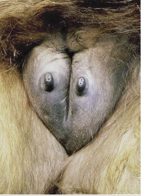
Figure 22-25 The udder is consolidated from right and left halves. The apices of the teats are perforated by the papillary ducts.
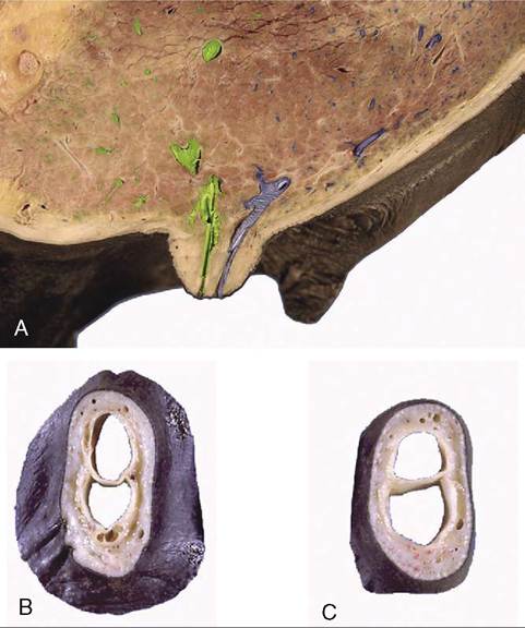
Figure 22-26 A, A sagittal section of the udder demonstrating the construction of the teat and the location of the lactiferous sinus. B, C, Transected teats showing internal division.
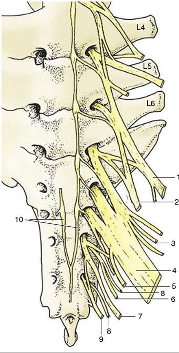
Figure 22-27 Ventral view of sacrum and caudal lumbar vertebrae with emerging ventral rami forming the lumbosacral plexus. 1, Femoral n., 2, obturator n., 3, cranial gluteal n. 4, sciatic n. 5, caudal cutaneous femoral n. 6, caudal gluteal n. 7, pudendal n. 8, pelvic n. 9, caudal rectal n. 10, continuation of sympathic cord.
nerve; the contributing spinal nerves are thus those of cord segments L2-4 and S2-4 (Figure 22-27). The substance of the gland is supplied by the genitofemoral nerve (L3-4). The glands develop rapidly during the second half of the first pregnancy and commence secretion before birth. Sebaceous secretion, epithelial debris, and possibly colostrum that escape through the teat openings during the last days of pregnancy dry to give the apex a waxy covering, which is a useful indication that parturition impends.