The Urethra and Accessory Reproductive Glands (see also pp. 180-182)
The very short first part of the male dog's urethra is completely surrounded by the prostate (Fig. 15.17 and Fig. 5.1/9). It presents a lumen indented by a dorsal ridge, locally raised to form a seminal colliculus that is perforated to each side by the narrow opening of the deferent duct and the numerous pores that drain the prostate.
The remaining part of the pelvic urethra is provided with a thin sleeve of spongy tissue within the striated urethralis muscle. The urethral lumen widens caudal to the prostate but narrows again as it leaves the pelvis at the ischial arch. In the tom, the prostate is located 3 to 4 cm caudal to the bladder neck, and the preprostatic part of the urethra has sometimes been described as an elongated bladder neck. The striking radiographic appearance of the feline urethra is shown in Fig. 15.8/1-3.The ampullary glands and prostate provide the entire complement of accessory sex glands in the dog. In dogs, sometimes remnants of the paramesonephric duct (vagina masculine) are present in the genital fold, covered dorsally by the prostate.
The cat, which lacks ampullary glands, has small bulbourethral glands located on the urethra, level with the ischial arch. These glands are important landmarks in perineal urethrostomy (removal of the penis in chronic urethral obstruction). The pudendal nerve courses over the ventral part of the bulbourethral glands.
In both species the prostate contributes the bulk of the seminal fluid. In the dog, it comprises a large compact mass about the urethra and neck of the bladder and a small disseminate part spread within the urethral mucosa. The compact part varies greatly in size, and the variation obviously affects its position and relations. It may be within the pelvic cavity when small, but more usually, and especially in mature and older dogs, it is mainly if not entirely intraabdominal (Fig.
15.18/2). A dorsal groove and internal septum divide it into right and left lobes, which are subdivided into lobules by finer septa that radiate outward to the capsule. The right and ventral lobes do not join ventral to the urethra in cats.The prostate is extremely sensitive to hormonal influences, and it is difficult to suggest normal dimensions because hyperplasia of the parenchymatous part commonly develops in early middle age and fibrosis and shrinkage are common senile changes. The hyperplasia sometimes affects the different lobes unequally. An enlarged prostate may press on the large intestine, producing constipation and difficulties in defecation; however, in contrast to the human experience, interference with micturition is unusual unless the condition is very gross. The state of the prostate —its size, firmness, and regularity of form—may be assessed by digital examination per rectum, a procedure facilitated by pushing the bladder toward the pelvis with pressure applied through the abdominal wall. The proportions of parenchyma and supporting tissue may be estimated from gross sections of autopsy specimens: connective tissue normally predominates in the prostate of the very young, glandular tissue predominates in those from animals in their prime, and the relationship is inconstant in the glands of aged dogs. It has been reported that the prostate is proportionately much larger (by a factor of four) in the Scottish terrier than in other breeds.
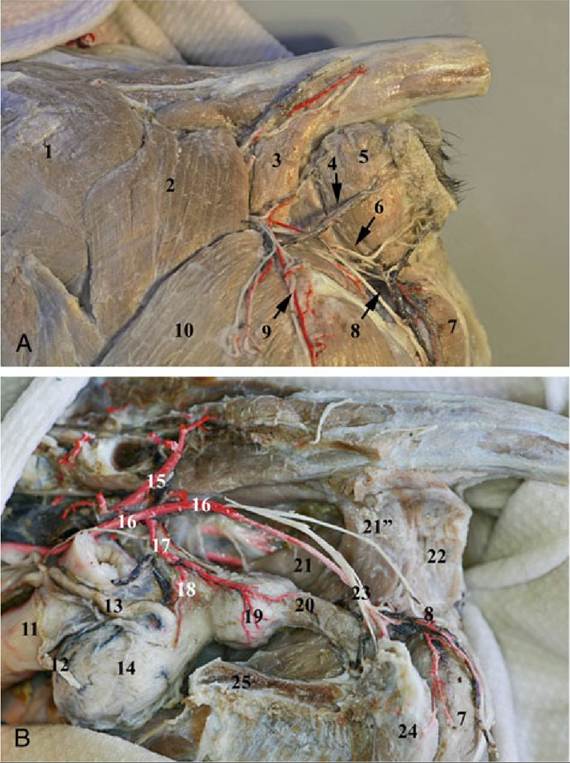
FIG. 15.17 (A) Gluteal and perineal region of the dog. (B) Pelvic cavity after removal of the rectum and the anus. 1, Gluteus medius muscle; 2, superficial gluteus muscle; 3, coccygeus muscle; 4, caudal rectal vessels and nerve; 5, sphincter ani externus muscle; 6, superficial perineal vessels; 7, bulbospongiosus muscle; 8, perineal nerve, cutaneous branch; 9, caudal cutaneus femoral nerve; 10, biceps femoris muscle; 11, rectum; 12, deferent duct; 13, ureter; 14, urinary bladder; 15, cranial gluteal artery; 16, internal pudendal artery; 17, prostatic artery; 18, caudal vesical artery; 19, prostate gland with the prostatic artery; 20, pelvic urethra with the urethral branch of the prostatic artery; 21, levator ani muscle (pars iliocaudalis);
21’, pars pubocaudalis; 22, superficial part of the sphincter ani externus muscle; 23, pudendal nerve (branches to the penis); 24, ischiocavernosus muscle; 25, symphysis pelvis (cut).
Enlargement of the prostate is sometimes treated by castration. Alternatively, or if castration fails, surgical removal may be performed. It is then relevant to note that generally only the craniodorsal aspect of the gland has a peritoneal covering. The trunk of the prostatic artery continues over the lateral aspect of the gland as the supply to the bladder after detaching prostaticovesical and prostaticourethral branches. The other structure at risk is the plexus formed by the pelvic and hypogastric autonomic nerves.
Beyond the prostate, the urethra widens before narrowing on leaving the pelvis and becoming incorporated in the penis. It is narrowest just before opening to the exterior at the tip of the glans, where urinary calculi, a frequent affliction of male cats, are often held up. Little is known of age changes to the prostate of this species, in which enlargement is a much less frequently encountered problem (Fig. 15.22/8).