The Penis and Prepuce (see also pp. 182-184)
The penis of carnivores has several unusual features, and additional differences between the organs of the dog and cat make separate description necessary.
The penis of the dog is slung between the thighs, where it may be palpated along its whole length.
The root is formed of two slender crura that arch forward from their ischial attachments to combine in a common body that is little stouter than either contributor (Fig. 15.19/4'). The urethra is incorporated at the same level and runs forward on the ventral surface of the body (Fig. 15.19/3). At the level of the ischial arch the corpus spongiosum (which there surrounds the urethra) expands to form the paired bulbus penis (covered by the bulbospongiosus muscle, a continuation of the urethralis muscle); further distally, the corpus spongiosum expands to form the glans penis, which is unusually extensive and clearly divided, both externally and internally, into a proximal expanded part (bulbus glandis; Fig. 15.19/7) and a distal cylindrical part (pars longa glandis; Fig. 15.19/7'), which provides the apex. About half the bulbus and the whole pars longa project into the preputial cavity, where they may be palpated. The cavernous parts of both crura combine within the proximal part of the body to form a single corpus cavernosum (Fig. 15.19/4) with a tough outer fibrous covering and a substantial median septum; these are connected by radial trabeculae that divide and enclose relatively meager cavernous spaces. The corpus cavernosum comes to a premature end because its distal part is converted into a bone, the os penis, within the core of the organ (Fig. 15.19/5). This bone is grooved ventrally for the reception and protection of the urethra within its spongy covering; the bone tapers toward its distal extremity, which is prolonged by a short, ventrally deflected rod of fibrocartilage that reaches almost to the very apex of the penis. The fibrocartilage remains unossified even in aged animals. The partial enclosure of the urethra within the groove of the os penis impedes the passage of urethral calculi, which therefore tend to lodge at the caudal end of the bone.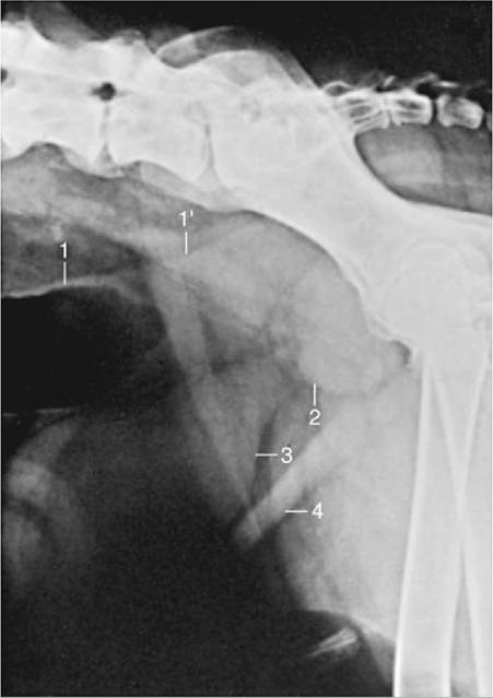
FIG. 15.18 Lateral radiographic view of the canine caudal abdomen to show the position of the prostate. 1 and 1', Descending colon containing gas and feces; 2, prostate; 3, bladder; 4, abdominal floor.
The caudal (or proximal) part of the glans penis, the bulbus glandis (Fig. 15.19D), is considerably expanded, even in the quiescent state. It is firmly anchored to the bone and considerably overlapped by the elongated distal division, which presents the urethral orifice toward its tip. The pars longa is more loosely attached to the bone. Both contain large blood spaces enclosed by relatively weak trabeculae.
The structure and connections of the various erectile bodies and their relationships to the supplying and draining vessels require close attention if the mechanism of erection is to be understood (Figs. 15.20 and 15.21). The penis is supplied by the continuation (beyond the origin of its perineal branch) of the internal pudendal artery, which now becomes the artery of the penis (Fig. 15.20/1'). The artery of the penis divides into three. One division, the artery of the bulb (Fig. 15.20/2), supplies the bulb (of the penis) and then runs distally within the organ to supply the corpus spongiosum about the urethra and later, on approaching the apex of the penis, the elongated portion of the glans. The second, the deep artery of the penis (Fig. 15.20/3), supplies several branches to both the tissues and the blood spaces of the corpus cavernosum. The third, the dorsal artery of the penis (Fig. 15.20/4), may be regarded as the direct continuation of the main trunk. It first runs on the dorsal aspect of the penis before sinking to the side and dividing close to the caudal limit of the bulbus.
A superficial branch runs almost to the tip of the organ below the skin over the ventral aspect of the glans; a deep branch penetrates the bulbus to run apically on the os penis to enter the pars longa; and a preputial branch forks into a division that runs over the dorsal aspect of the bulbus to supply the dorsal aspect of the pars longa and the prepuce.Internal pudendal artery
Artery of the penis
Artery of the bulb: Supplies the bulb, the corpus spongiosum, and the elongated part of the glans Deep artery of the penis: Supplies the tissue and the caverna of the corpus cavernosum
Dorsal artery of the penis: Supplies the caudal parts of the bulbus, the skin over ventral aspect of the glans, and the pars longa and prepuce
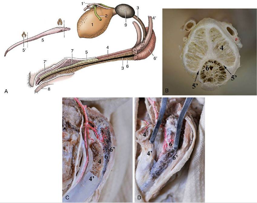
FIG. 15.19 Canine bladder, urethra, and penis (in section) schematic (A), transected (B), root of penis (C), and bulbus penis (D). 1, Bladder; 1', left ureter; 2, left deferent duct; 3, urethra; 4, corpus cavernosum; 4', left crus; 5, os penis; 5', urethral groove; 6, corpus spongiosum; 6', bulb of penis; 7, bulbus glandis; 7', pars longa glandis; 8, prepuce; 9, prostate.
The veins are broadly satellite to the arteries. The dorsal vein leaves the lateral aspect of the bulbus and runs caudally, gradually shifting toward the dorsal aspect of the penis, where it is joined by a common trunk formed of the veins corresponding to the deep artery and the artery of the bulb. The augmented dorsal vein then bends around the ischial arch to enter the pelvis, where it provides the main radicle of the internal pudendal vein. Other veins assist in the drainage of the glans. A superficial vein leaves the pars longa to wind around the fornix of the prepuce before joining the external pudendal vein. A deep vein within the glans drains blood from the pars longa to the bulbus; it is valved so that reflux of blood is impossible and is so arranged that it may either provide a through passage to the dorsal vein or open into the blood spaces of the bulbus, from where the blood then enters the dorsal vein.
The usual muscles are present. The retractor penis, largely composed of smooth muscle, loops to the side of the anal canal before converging on its fellow to form a band that runs along the urethral aspect of the penis to a termination by the preputial fornix. A few small fascicles are detached to the scrotum. Short but powerful ischiocavernosus muscles cover the crura. The bulbospongiosus forms a transverse covering over the urethra from the bulb to its incorporation in the penis. A small ischiourethralis passes from the ischial tuber to a fibrous ring that encloses the dorsal veins at their entry to the pelvis. The two large muscles at the root of the penis can be identified on palpation (Fig. 15.3/6 and 7).
The prepuce of the dog is rather pendulous toward its cranial extremity, where it is suspended below the abdomen by a fold of skin. It has a simple arrangement, and the parietal part of its lining is studded with lymph nodules, which give it a rather irregular appearance. There are also small scattered preputial glands. Paired preputial muscles, detachments from the cutaneous muscle of the trunk, run over the abdominal floor to meet and partially decussate in the skin of the prepuce caudal to the T-shaped orifice.
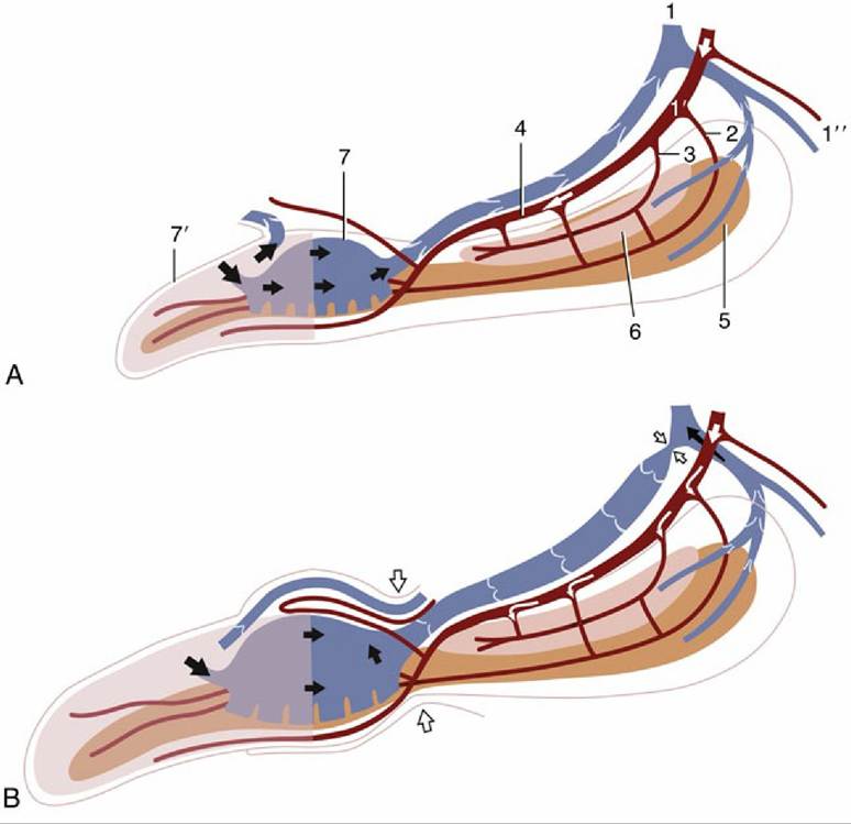
FIG. 15.20 Schematic representation of the blood supply and the blood spaces of the (A) quiescent and
(B) erect canine penis. 1, Internal pudendal vessels; 1', artery of the penis; 1", perineal branches; 2, artery of the bulb; 3, deep artery of the penis; 4, dorsal artery of the penis; 5, corpus spongiosum; 6, corpus cavernosum; 7, bulbus glandis; 7', pars longa glandis.
Phimosis and Paraphimosis: Congenital or acquired narrowing of the preputial orifice is rare and may prevent protrusion of the penis (phimosis). Those acquired cases that are due to scar formation after an earlier inflammation may be treated surgically. The wisdom of surgical intervention may be questioned when the defect is congenital and possibly hereditary.
Paraphimosis, in which the erect penis is unable to subside and cannot be withdrawn into the prepuce, requires more urgent attention because the interruption of the circulation may cause tissue death within hours. The surgical intervention, called phallopexy, is required to create an attachment between the shaft of the penis and the mucosa of the prepuce.At birth, the epithelial surface of the prepuce and penis adhere through a frenulum. Separation of the prepuce from the penis is under androgenic influence and usually occurs at puberty.
The penis of the cat is unique (among domestic species) in retaining the embryonic position: the apex is directed caudoventrally and the urethral surface is uppermost (Figs. 15.22/6 and 15.23). Relatively much shorter than the penis of the dog, that of the cat has a similar construction, including the transformation of the distal part of the corpus cavernosum into bone. Kittens lack the os penis until 3 months of age. The existence of an apical ligament extending between the os penis and the proximal part of the corpus cavernosum appears to be responsible for the ventral deflection of the penis that occurs with erection. The dorsal artery supplies only the prepuce and not the penis. The glans is small, and its free surface is generously ornamented with small, keratinized spines in the tom; these spines develop during the first few months of postnatal life and regress to a very insignificant state in castrated animals (Figs. 15.23 and 15.24). Approximately 120 in number, they lie flat against the surface of the glans in the nonerect state but rise, as a result of the congestion of the blood spaces at their bases, on erection. The stimulus they provide to the queen is believed to be important in inducing ovulation.
The cat's prepuce is thick but short and often much obscured by hair; its orifice faces caudally, and urine is ejected in this direction. The spraying of urine by the tom is a social gesture marking territory (Fig. 15.25). The sites are not always discretely chosen and are often inconvenient to the owner, one reason for the common practice of castration.*
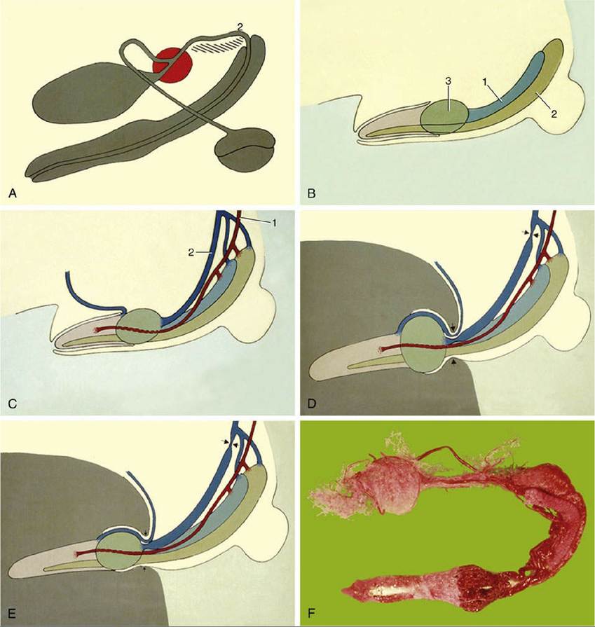
FIG. 15.21 (A) Schematic representation of canine male reproductive organs. (B) Major vascular parts of canine penis. 1, Corpus cavernosum; 2, corpus spongiosum; 3, bulbus glandis. (C), (O), and (E) Stages in the erection process. 1, Penile artery; 2, dorsal penile vein. (F) Corrosion cast of the arterial supply to the prostate and penis.