THE UTERUS
The uterus, the womb in popular speech, is the enlarged part of the tract in which embryos come to rest, where they establish a means of physiological exchange with the mother’s bloodstream, and where they are protected and nourished until ready to be delivered to the outside world.
It is the part of the tract that displays the most striking specific differences, although the most extreme forms do not occur among domestic species. These differences find a ready explanation in the manner of formation of the reproductive tract (p. 172) from two paramesonephric ducts that grow caudally to meet and fuse with each other and with the median urogenital sinus, the ventral division of the cloaca (see Figures 5-15 and 5-16). In some species, including many rodents, fusion of the ducts is limited to the most caudal portions, which contribute to the vagina; the more cranial parts remain distinct, and the uterus thus consists of paired tubes that open separately into the vagina (double uterus—uterus duplex). In contrast, in women and most other primates, fusion is much more extensive and only the uterine tubes remain paired; a median uterus with a simple undivided lumen is present. In the intermediate variety (bicornuate uterus) found in all major domestic species, the uterus comprises a caudal median part from which paired horns diverge cranially to continue as the uterine tubes.In all domestic mammals the median part of the uterus has two segments. The caudal, very thick-walled segment, the cervix (Figure 5-59/8), provides a sphincter controlling access to and from the vagina. A part of the cervix (Figure 5-59/9) (portio vaginalis) usually projects into the vaginal lumen with which it communicates at the external ostium. The lumen of the cervix (cervical canal) is constricted and often almost occluded by mucosal folds; it opens into the body of the uterus (Figure 5-59/6) at the internal ostium.
The body is generally a rather small segment in domestic species, although the proportions vary (see Figure 5-16); it is largest in the mare. The division of the interior is not always obvious externally because an internal septum may partially divide an apparently single space. Although visual inspection generally fails to reveal the extent of the cervix, this is easily discovered on rectal palpation as it is much firmer than the adjacent parts.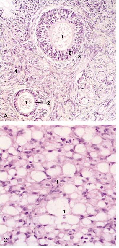
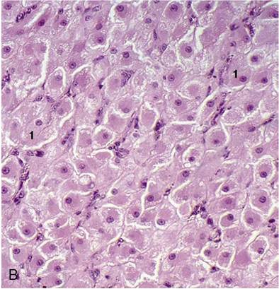
Figure 5-57 A, Ovary (bitch) in anestrus with preantral follicles (140?). 1, oocyte; 2, granulosa cells; 3, theca cells; 4, stroma. B, Active corpus luteum (queen) (140?). C, Inactive corpus luteum (queen) (140?). 1, Degenerating luteal cells.
The horns (cornua) vary greatly in length, and it is hardly surprising that they are longest in polytocous species. Their disposition also varies; they are characteristically wound in ruminants, straight and divergent in mares and bitches, and cast into intestine-like loops in sows. The cervix generally lies within the pelvic cavity, interposed between the rectum and the bladder (Figure 5—32/7), but the body and horns of the uterus typically lie within the abdomen above the mass of intestines.
The uterus possesses serosal, muscular, and mucosal coats that are known as the perimetrium, myometrium, and endometrium, respectively. The serosal covering reaches the uterus by extension from the supporting broad ligament (mesometrium; Figure 5-33/7). The muscle is arranged as weak external longitudinal and thicker internal circular layers that are separated by a very vascular stratum of connective tissue. The tissues, especially the external muscle layer, extend (as parame- trium) into the supporting broad ligaments. Dense connective tissue intermingles with the muscle of the cervix and makes this a very indistensible part of the tract at most times (Figure 5-60).
The endometrium is thick. Its surface relief varies among species and is most remarkable in ruminants, in which numerous permanent elevations (caruncles) mark the sites where the embryonic membranes firmly attach during pregnancy (Figure 5-59/7). Numerous tubular glands open on the surface, which is generally lined by a simple columnar epithelium. The mucosa within the cervix is prominently modeled by both longitudinal and circular folds whose interdigitation helps close the passage (Figure 5-59/8). Mucus secreted by cervical glands plugs the canal at most times and so helps seal the uterus from the vagina. The passage is open only at estrus and immediately before, during, and, for a short time, after parturition.
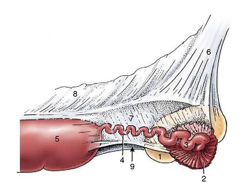
Figure 5-58 Lateral view of the suspension of the right ovary, uterine tube, and uterine horn of a mare. 1, Ovary; 2, infundibulum of tube; 3, ampulla of tube; 4, isthmus of tube; 5, uterine horn; 6, mesovarium; 7, mesosalpinx; 8, mesometrium; 9, arrow indicates entrance to ovarian bursa.
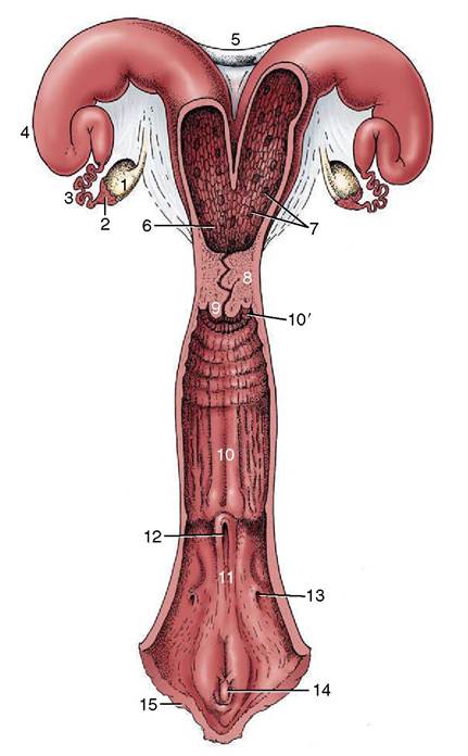
Figure 5-59 The reproductive tract of a cow, opened dor- sally. 1, Ovary; 2, infundibulum; 3, uterine tube; 4, horn of uterus; 5, intercornual ligaments; 6, body of uterus; 7, caruncles; 8, cervix; 9, vaginal part of cervix; 10, vagina; 10, fornix; 11, vestibule; 12, external urethral opening; 13, opening of major vestibular gland; 14, clitoris; 15, vulva.