The Uterus (see also pp. 186-187)
The uterus, which lies mainly dorsal to the small intestine, consists of a very short (about 2 to 3 cm) body and two long and slender (about 12 ? 1 cm) divergent horns (Figs. 15.11/7 and 8 and 15.12).
The body is near the pubic brim but may be abdominal or pelvic in position. It is, in fact, even shorter than external inspection suggests because a short internal septum continues caudally from the junction of the horns. The cervix is also very short—the canal is barely 1 cm long—but the tissue thickening extends beyond the external ostium as a fold on the roof of the vagina (Fig. 15.12/3 and 3'). Transverse grooves frequently divide this fold into cranial, middle, and caudal tubercles; they become very swollen at certain stages of the cycle. The ostium of the cervix generally faces caudoventrally, and this orientation, combined with the asymmetry of the fornix and the fissuration of the cervical prolongation, may make its identification rather difficult, even with the aid of an endoscope.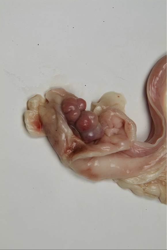
FIG. 15.10 Ovarian bursa opened to expose the ovary (bitch).
The feline cervix feels like a hard oval knot at the uterovaginal junction and, although small, is readily distinguished from the adjoining parts by the thickness of its wall. As in the bitch, the cervical mucosa is smooth, without conspicuous folds.
The broad ligaments also commonly contain much fat. They are wider in their middle parts than toward their extremities and allow the horns of the uterus considerable mobility. An unusual feature is the detachment from the lateral surface of a peritoneal fold that extends toward, and in the bitch through, the inguinal canal to end variously between the groin and the vulva. The fold is thickened at its free margin (the round ligament), slightly dilating the canal and predisposing to inguinal hernia, an almost male prerogative in other species.
The most likely herniation is of the uterine horn, which may result in its subcutaneous entrapment of the pregnant uterus and may require a separate procedure to deliver the fetus.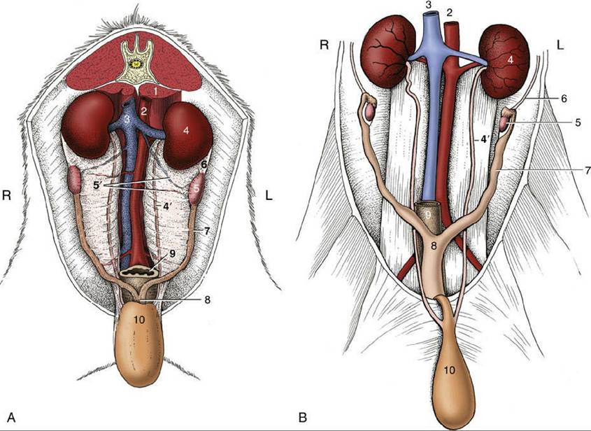
FIG. 15.11 (A) Canine and (B) feline ovaries and uterus in situ, ventral view. 1, Psoas muscles; 2, aorta; 3, caudal vena cava; 4 and 4', left kidney and ureter; 5, ovary; 5', ovarian vessels; 6, suspensory ligament of ovary; 7, uterine horn; 8, body of uterus; 9, rectum; 10, bladder, reflected caudally; L, left; R, right.
The vascularization of the uterus depends on the uterine branch of the ovarian artery and the uterine artery, a branch of the vaginal artery (Fig. 15.13/1 and 5). The two vessels anastomose within the broad ligament and must be ligated when ovariohysterectomy is performed. These vessels lie close to the extremities of the uterus but swing away in the intermediate part of the broad ligament. The proximity of the uterine artery to the cervix allows an arterial ligature to be securely anchored to the uterine stump to prevent slippage when the bulk of the uterus is removed surgically. The uterus is drained by left and right uterine veins that empty into the renal vein and the caudal vena cava, respectively. The ovarian artery and vein do not closely accompany each other within the mesovarium.
The lymphatic drainage of the ovary and uterus passes to the medial iliac and aortic lumbar nodes.