THE UTERUS (See also pp. 199-201.)
The uterus, which lies mainly dorsal to the small intestine, consists of a very short (ca. 2- to 3-cm) body from which two long and slender (ca. 12- ? 1-cm) horns diverge (Figure 15-10Z7,the uterine stump to prevent slippage when the bulk of the uterus is removed surgically.
Almost the entire uterus is drained by a large uterine tributary of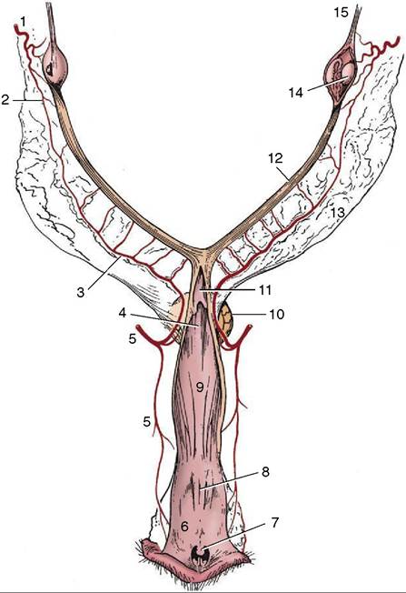
Figure 15-12 Blood supply of the reproductive organs of the bitch, dorsal view. The right ovarian bursa and the caudal parts of the tract have been opened. 1, Ovarian artery; 2, uterine branch of ovarian artery; 3, uterine artery; 4, dorso- median fold continuing the cervix; 5, vaginal artery; 6, vestibule; 7, clitoris; 8, external urethral orifice; 9, vagina; 10, bladder; 11, cervix; 12, right uterine horn; 13, broad ligament; 14, right ovary; 15, suspensory ligament of ovary.
the ovarian vein, which empties into the renal vein on the left but generally proceeds directly to the caudal vena cava. The ovarian artery and vein do not closely accompany each other within the mesovarium.
The lymphatic drainage of the ovary and uterus passes to the medial iliac and aortic lumbar nodes.