THE OVARIES AND UTERINE TUBES
(See also pp. 197-199.)
(Figure 15-8), which is a firm, flattened, ellipsoidal structure measuring about 15 ? 10 ? 6 mm. Its contours are obviously less regular in phases of the estrous cycle in which large follicles or corpora lutea are present (Figure 15-9).
The wall of the ovarian bursa of cats commonly contains conspicuously less fat than that of the bitch and covers only the lateral surface of the ovary, which is consequently more immediately visible.The ovaries (within the bursae) lie close to, or even in contact with, the caudal poles of the kidneys; in conformity with the asymmetrical position of the kidneys the left ovary is placed a little caudal to its fellow. Although most spays (the removal of ovaries and [parts of] the uterine horns; ovariectomy/ovariohys- terectomy) are now performed by midline incision, an alternative lateral approach is quite often used in cats. The flank incision is made midway between the iliac crest and the last rib in the confident expectation that
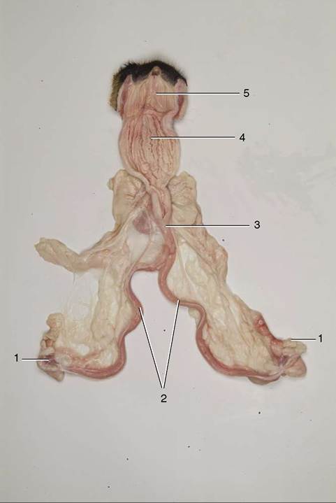
Figure 15-8 Overview of canine female reproductive tract. Vagina has been opened. 1, Ovaries; 2, uterine horns; 3, uterus body; 4, vagina; 5, vestibulum.
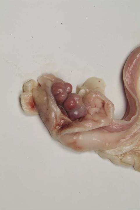
Figure 15-9 Ovarian bursa opened to expose the ovary (bitch).
the ovary will be within easy reach. The right ovary is usually found dorsal or dorsolateral to the ascending colon, and the left one is found between the dorsal extremity of the spleen and the descending colon. Lengthening of the attachments in parous animals allows the ovaries a greater mobility.
The ovary is fixed additionally by suspensory and proper ligaments. The former is a peritoneal fold, thickened along its free margin, that attaches to the transverse fascia close to the last rib in the dog (Figure 15-10/d); it is prolonged caudally as the proper ligament, which extends beyond the ovary to merge with the tip of the uterine horn. The anchorage provided by the suspensory ligament makes surgical exteriorization of the ovary difficult.
The suspensory ligament in the cat reaches the diaphragm and allows the ovary greater mobility.The entrance to the canine bursa is reduced to a slit in the medial wall, usually made obvious by the protrusion of a few reddish infundibular fimbriae. The infundibulum is continued by the narrower part of the uterine tube, which is not obviously divided between ampulla and isthmus. These parts follow a tortuous course within the walls of the bursa; disregarding minor kinks and bends, the tube runs in a broad sweep that first passes forward in the distal mesovarium before crossing cranial to the ovary to continue caudally in the mesosalpinx (Figure 5-60). It ends in an abrupt junction with the horn of the uterus. Although in most subjects much of the tube is concealed by fat deposits, the terminal part is usually visible. The infundibulum may transmit bacteria into the bursa (or abdominal cavity) in the case of pyometra.
Parovarian cysts originate from remnants of either mesonephric or paramesonephric ducts. They are more frequently encountered during ovariohysterectomy in dogs than in cats and are located between the ovary and uterine horn.
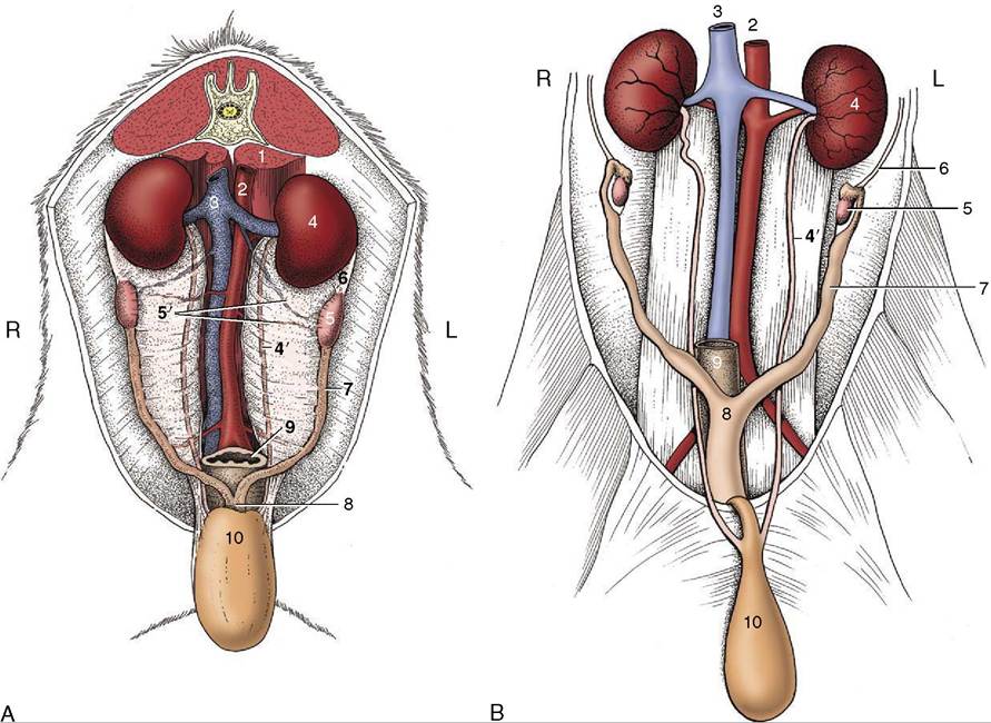
Figure 15-10 Canine (A) and feline (B) ovaries and uterus in situ, ventral view. 1, Psoas muscles; 2, aorta; 3, caudal vena cava; 4,4', left kidney and ureter; 5, ovary; 5', ovarian vessels; 6, suspensory ligament of ovary; 7, uterine horn; 8, body of uterus; 9, rectum; 10, bladder, reflected caudally.