THE VAGINA
The remaining part of the genital tract is divided between the vagina and vestibule, approximately in the ratio of 3:1; the boundary is a few centimeters cranial to the ischial arch (see Figure 29-11).
Because the vagina is capable of great expansion, in length and in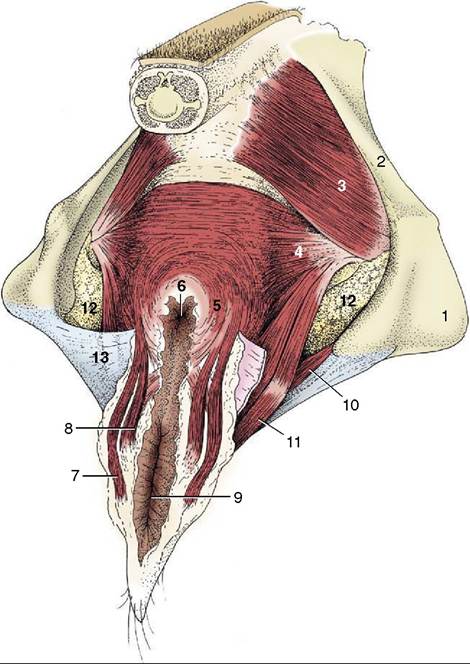
Figure 29-10 The perineal muscles of a cow. 1, Ischial tuber; 2, sacrosciatic ligament; 3, coccygeus; 4, levator ani; 5, external anal sphincter; 6, anus; 7, retractor clitoridis; 8, constrictor vulvae; 9, vulva; 10, urogenital diaphragm; 11, constrictor vestibuli; 12, fat in ischiorectal fossa; 13, perineal fascia (partly removed on the right side).
diameter, its passive dimensions are not of great significance. The lining exhibits low folds, both circular and longitudinal, and the lumen is closed by the falling together of the roof and floor (see Figure 29-8).
It is usual to find the caudal part ventrally narrowed, especially in young animals; this feature, not to be confused with the hymen (which is rarely much in evidence), is ascribed to the urethralis muscle.
The cranial two thirds of the dorsal wall faces into the rectogenital pouch, but caudal to this the vagina and rectum are joined by a wedge of tissue (see Figure 2911). The ventral surface has a less complete peritoneal covering and is related to the bladder and urethra and to the packing tissues about the urethra. The lateral walls are also largely without peritoneum, being crani- ally included in the broad ligament and more caudally sharing in the general retroperitoneal arrangement (see Figures 29-7 and 29-8). This limitation of the perito-
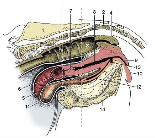
Figure 29-11 Median section of the bovine pelvis. The two vertical broken lines indicate the levels of the transverse sections in Figures 29-7 and 29-8. The position of the obturator foramen is indicated by a broken outline.
1, Sacrum; 2, first caudal vertebra; 3, rectum; 4, anal canal; 5, right uterine horn; 6, left uterine horn, mostly removed; 7, cervix; 8, vagina; 9, vestibule; 10, vulva; 11, bladder; 12, urethra; 13, suburethral diverticulum; 14, symphysis.
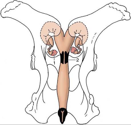
Figure 29-12 Dorsal view of the bony pelvis and related (nongravid) bovine reproductive organs. Note the position of the ovaries in relation to the pecten pubis. 1, Ovary; 2, cervix.
neum is relevant to the prognosis of wounds to the vaginal wall. The peritoneal covering of the dorsal fornix region provides a convenient route for surgical access to the abdominal cavity, most often used for operations on the ovary; it has the additional advantage

Figure 29-13 Various functional stages of the bovine ovary. A, Ovaries with small secondary follicles. B, Ovaries with mature follicle ready to rupture. C, Ovary with recently ruptured follicle; the scar is small and round. D, Ovary with mature corpus luteum.
of avoiding the major vessels that pass below and to the sides of the vagina (see Figure 26-19).
Vestiges of the mesonephric ducts may be found below the mucosa of the floor near the junction with the vestibule; they are sometimes the origin of cysts.
The vagina is almost absent in the freemartin (p. 712), whose abnormally short tract is evident on examination of the vestibule. Aplasia or constriction of the vagina also occurs in white heifer disease, another congenital anomaly.
The freemartin is found after a twin pregnancy in which the female fetus is adversely affected by the male twin (Figure 29-18).