The Vagina
The remainder of the female tract, although sometimes loosely termed the vagina, consists of two parts. The cranial part, the vagina in the strict sense (Fig. 5.59/10), is a purely reproductive passage that runs from the cervix to the entrance of the urethra.
The caudal part, the vestibule, extends from the urethral orifice to the external vulva and combines reproductive and urinary functions. The two parts together constitute the female copulatory organ and birth canal.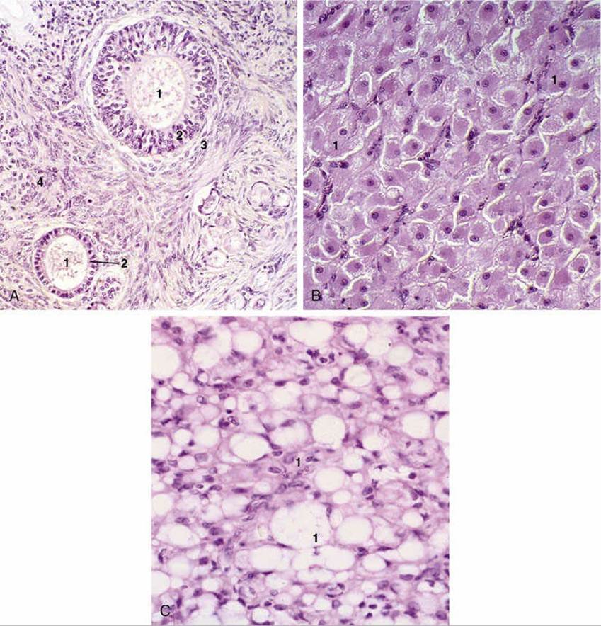
FIG. 5.57 Histologic preparations. (A) Ovary (bitch) in anestrus with preantral follicles (140?). 1, Oocyte;
2, granulosa cells; 3, theca cells; 4, stroma. (B) Active corpus luteum (queen) (140?). (C) Inactive corpus luteum (queen) (140?). 1, Degenerating luteal cells.
The vagina is a relatively long, thin-walled tube that is distensible in length and width. It occupies a median position within the pelvic cavity, related to the rectum dorsally and the bladder and urethra ventrally (Fig. 5.32/8). It is mostly retroperitoneal, although peritoneum does cover the cranial parts of both the dorsal and the ventral surfaces to a variable extent. Incision of this part of the dorsal wall, a relatively easy procedure to perform from within the vagina in larger species, provides a convenient access to the peritoneal cavity (see Fig. 22.6/2 and 8). The corresponding ventral approach is prohibited by the presence of a plexus of veins draining the uterus and vagina.
The vaginal muscle, although weaker, has a similar disposition to that of the uterus. The mucosa is lined by a stratified squamous epithelium that reacts, more emphatically in some species than in others, to changes in hormone levels throughout the estrous cycle. Glands are confined to the cranial part of the vagina, although the moisture may diffuse more widely.
The surface is smooth but circular, and longitudinal folds may form when the walls of the inactive organ collapse inward. The intrusion of the cervix into the cranial part of the vagina reduces the lumen of this part to a (generally) ringlike space known as the fornix (Fig. 5.59/10').The junction of vagina and vestibule is supposedly marked in virgin animals by a transverse mucosal fold (hymen). This is best developed in the filly and the gilt, but even in these it is rarely very prominent. It does not survive coitus. The junctional region is less distensible than the parts of
the tract cranial and caudal to it.
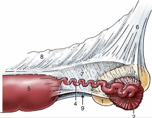
FIG. 5.58 Lateral view of the suspension of the right ovary, uterine tube, and uterine horn of a mare. 1, Ovary; 2, infundibulum of tube; 3, ampulla of tube; 4, isthmus of tube; 5, uterine horn; 6, mesovarium; 7, mesosalpinx; 8, mesometrium; 9, arrow indicates entrance to ovarian bursa.
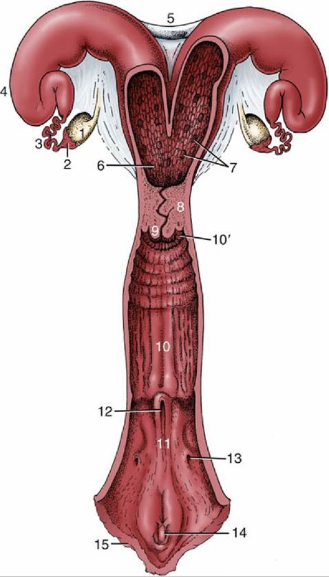
FIG. 5.59 The reproductive tract of a cow, opened dorsally. 1, Ovary; 2, infundibulum; 3, uterine tube; 4, horn of uterus; 5, intercornual ligaments; 6, body of uterus; 7, caruncles; 8, cervix; 9, vaginal part of cervix;
10, vagina; 10', fornix; 11, vestibule; 12, external urethral opening; 13, opening of major vestibular gland; 14, clitoris; 15, vulva.