The Veins
The two cranial venae cavae (Fig. 37.16/7) are satellite to the brachiocephalic arteries and receive tributaries (jugular and subclavian veins) from the neck and head and the breast and wing.
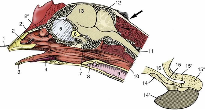
FIG. 37.38 Median section of the head with an enlargement of the hypophysis (inset). The arrow indicates the approach to the foramen magnum through which euthanasia may be performed by injection into the brain. 1, Wire in nostril; 2, 2', and 2", rostral, middle, and caudal nasal conchae, respectively; 3, mandible; 4, tongue; 5, interorbital septum; 6, optic chiasm; 7, hypophysis (see also inset); 8, larynx; 9, trachea; 10, esophagus; 11, spinal cord; 12, cerebellum; 13, cerebrum; 14 and 14', pars tuberalis and pars distalis of the adenohypophysis, respectively; 15, 15', and 15", median eminence, infundibulum, and neural lobe of the neurohypophysis, respectively; 16, third ventricle.
Venipuncture: The right jugular vein, always larger than the left, is visible through the skin and available for venipuncture (Fig. 37.15/6). However, this is not possible in pigeons, in which the skin is very thick in this area. Venipuncture in these birds is done from the medial metatarsal vein. In many small cage birds the left jugular is very small. The cutaneous ulnar vein (wing vein), subcutaneous on the ventral surface of the extended wing, may also be used for the administration of fluids or collection of very small volumes of blood (Fig. 37.13/9). The habit of clipping a claw for a small amount of blood is now condemned: it is much better to puncture the medial metatarsal vein.
The caudal vena cava drains the liver, kidneys, gonads, and oviduct. It forms ventral to the kidneys from the union of the common iliac veins that drain the pelvis and hindlimbs (Fig. 37.29/13). As described on page 791, some blood from the pelvis and hindlimbs passes through the kidneys (renal portal system) before reaching the caudal vena cava. Blood from the gastrointestinal tract reaches the liver by separate right and left hepatic portal veins that enter the respective lobes. The left vein drains the left and ventral parts of the stomach. The much larger right vein drains the right and dorsal parts of the stomach, the spleen, and the remainder of the gastrointestinal tract through cranial and caudal mesenteric veins. The caudal mesenteric vein, connected to the caudal end of the renal portal system (Fig. 37.29/22), also conveys a considerable amount of blood toward the kidneys. Thus, some blood from the gastrointestinal tract may return to the heart without passing through the liver.